RSNA 2019 Presents Session on Lung Injury from Vaping
Released: December 02, 2019
- RSNA Media Relations
1-630-590-7762
media@rsna.org - RSNA 2019 Newsroom
(Dec. 1-5, 2019)
1-312-791-6610 - Linda Brooks
1-630-590-7738
lbrooks@rsna.org - Dionna Arnold
1-630-590-7791
darnold@rsna.org
CHICAGO — A panel of medical professionals will discuss the public health impact of e-cigarette use, or "vaping," today during a session at the annual meeting of the Radiological Society of North America (RSNA).

Mark L. Schiebler, M.D.

Jeffrey S. Klein, M.D.
E-cigarette use is on the rise. According to the Centers for Disease Control and Prevention (CDC), more than 9 million adults in the U.S. use e-cigarettes, and vaping has become especially popular among teens. The 2018 National Youth Tobacco Survey reported that in 2018 more than 3.6 million middle and high school students were using e-cigarettes.
E-cigarette inhalants, upon vaporization of the e-cigarette solution, contain potentially harmful toxic substances that can cause lung injury and, in some cases, inhibit vascular function.
In August, a multistate outbreak of lung disease associated with e-cigarette product use began in the United States. The situation is ongoing, and multiple substances and product sources are still under investigation by the CDC. However, most patients involved in the outbreak have reported using products containing tetrahydrocannabinol (THC). Therefore, the CDC recommends that people avoid e-cigarette products that contain THC.

Brandon Larsen, M.D., Ph.D.

Seth J. Kligerman, M.D.
"An outbreak of lung disease associated with vaping has developed recently throughout the U.S.," said Jeffrey S. Klein, M.D., RSNA Board Liaison for Publications and Communications and A. Bradley Soule and John P. Tampas Green and Gold Professor of Radiology at the University of Vermont College of Medicine in Burlington. "Radiographic and CT findings of diffuse pneumonitis have emerged as characteristic findings in affected patients and are included in the CDC case definition of vaping-induced lung injury.
"In our efforts to educate radiologists about this potentially fatal condition they may encounter in younger patients with acute respiratory symptoms, the RSNA has organized a special session at RSNA 2019 where experts will provide up-to-date information regarding key radiologic, pathologic and physiologic findings associated with this critical public health issue," he said.
Dr. Klein participated in calls with the CDC, as it worked with various medical specialty groups to investigate and identify potential causes of vaping-related lung injury. In September, Dr. Klein produced a brief educational video aimed at updating radiologists on this topic, so that they could be well positioned to assist in identifying cases.

Alessandra Caporale, Ph.D.
For the session at RSNA 2019, Dr. Klein will be joined by Mark L. Schiebler, M.D., professor of cardiothoracic radiology at University of Wisconsin School of Medicine and Public Health - Madison, Travis S. Henry, M.D., associate professor of clinical radiology at the University of California, San Francisco, Seth J. Kligerman, M.D., associate professor of radiology and section chief of cardiothoracic imaging at the University of California, San Diego, Brandon Larsen, M.D., Ph.D., consultant and associate professor of laboratory medicine and pathology at Mayo Clinic in Scottsdale, Ariz., and Alessandra Caporale, Ph.D., post-doctoral researcher in the Laboratory for Structural, Physiologic and Functional Imaging at the University of Pennsylvania Perelman School of Medicine in Philadelphia.
The session will provide a brief introduction, followed by an exploration of the scope of the problem, description of imaging findings and histopathology associated with vaping-related lung injury, a presentation on how vaping affects the vascular system and a Q&A with the panelists.
After attending the session at RSNA 2019, radiologists and other medical professionals should have a greater understanding of the public health implications of the vaping-related lung injury outbreak in the U.S. They will also become familiar with common CT and X-ray findings associated with the condition, and how the pathology helps to define the possible causes of this disorder. Lastly, they will know more about vaping's impact on vascular function.
"As we learn more, RSNA will continue to keep radiologists informed on this important public health topic," Dr. Klein said.
"Special Interest: E-cigarette/Vaping-associated Lung Injury (EVALI)" (SPSI27) will be held Monday, Dec. 2, from 4:30 – 5:30 p.m.
For the latest information from the CDC on vaping-associated lung injury, visit https://www.cdc.gov/tobacco/basic_information/e-cigarettes/severe-lung-disease.html.
Note: Copies of RSNA 2019 news releases and electronic images will be available online at RSNA.org/press19 beginning Monday, Nov. 25.
RSNA is an association of over 53,400 radiologists, radiation oncologists, medical physicists and related scientists, promoting excellence in patient care and health care delivery through education, research and technologic innovation. The Society is based in Oak Brook, Ill. (RSNA.org)
To ensure you are using the most up-to-date information, please call the RSNA Newsroom at 1-312-791-6610.
For patient-friendly information on chest X-ray and CT, visit RadiologyInfo.org.
Images (JPG, TIF):
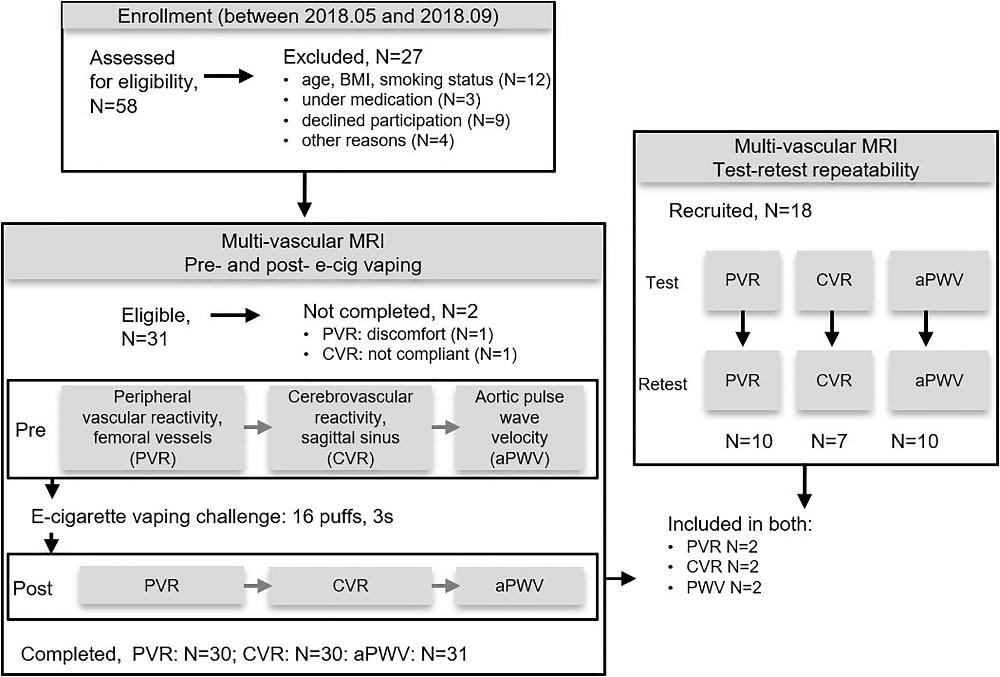
Figure 1 (Caporale). Flowchart of participant enrollment and exclusion, completed assessments, and test-retest repeatability. aPWV = aortic pulse wave velocity, BMI = body mass index, CVR = cerebrovascular reactivity, PVR = peripheral vascular reactivity.
High-res (TIF) version
(Right-click and Save As)

Figure 2 (Caporale). Response to cuff occlusion in the femoral circulation. A, Vessel-wall images of the superficial femoral artery (SFA) at different points (60 seconds [A60], 90 seconds [A90], and 120 seconds [A120]) as indicated by crosses in B during reactive hyperemia. The dashed circles represent the lumen area at baseline. B, Superficial femoral vein (SFV) oxygen saturation (SvO2) at baseline (green line) and during hyperemia. C, SFA blood flow velocity (V). D, Axial image obtained with MRI in the thigh, with SFA and SFV indicated in red and blue, respectively. Sample data shown for a representative participant. ΔSvO2 = peak-to-peak SvO2, HI = hyperemic index, PFR = peripheral flow reserve, RI = resistivity index, TFF = time of forward flow, TP = time to peak, TW = washout time, Vb = baseline velocity, VP = peak hyperemic velocity, Vr = retrograde velocity during early diastole, Vs = systolic velocity.
High-res (TIF) version
(Right-click and Save As)
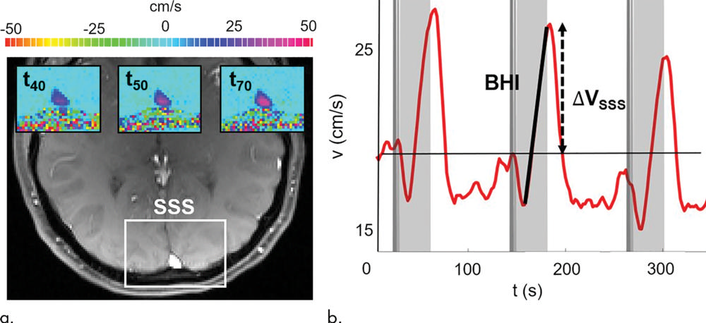
Figure 3 (Caporale). Neurovascular response to breath hold. (a) Magnitude image intensity of superior sagittal sinus (SSS, box). Insets show velocity maps at different points of the velocity time-course (40 seconds [t40], 50 seconds [t50], and 70 seconds [t70]). (b) Sample SSS blood flow velocity time-course (red line) shown for a representative participant. The thick black line is linear fit during breath holds, the slope of which is the breath-hold index (BHI). ΔVSSS = post–breath hold relative velocity increase.
High-res (TIF) version
(Right-click and Save As)
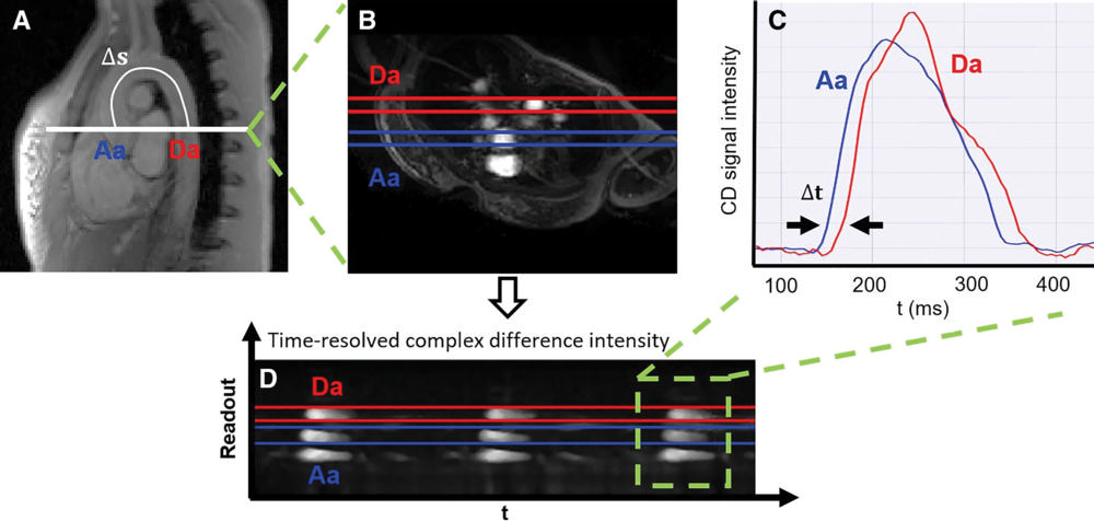
Figure 4 (Caporale). Measurement of aortic pulse wave velocity. A, Sagittal view of the aortic arch with path length of the flow wave between the ascending aorta (Aa) and descending aorta (Da; Δs). B, Axial view showing Aa and Da, and the time course of the, C, complex difference (CD) signals during three cardiac cycles. D, CD signal intensity averaged across the aorta width during a single cardiac cycle (the transit time [t] is indicated). Sample data shown for a representative participant. t = time.
High-res (TIF) version
(Right-click and Save As)
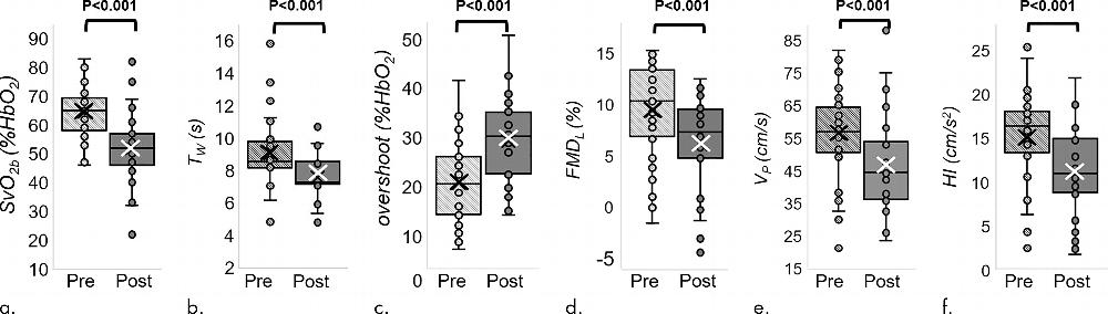
Figure 5 (Caporale). MRI-derived vascular parameters before and after e-cigarette vaping. Box-and-whisker plots for the strongest effects observed show (a) SvO2 during baseline (SvO2b ), (b) SvO2 washout time (TW), (c) SvO2 overshoot, (d) luminal flow-mediated dilation (FMDL), (e) peak hyperemic blood flow velocity (VP), and (f) hyperemic index (HI). P values were derived on the basis of paired t tests. The boxes represent inner quartiles; horizontal lines within the box indicate the median and crosses (X) indicate the mean.
High-res (TIF) version
(Right-click and Save As)

Figure 6 (Caporale). Electronic cigarette components. Photograph shows the superior envelope (1), mouthpiece (2), light emitting diode (3), cylindrical 3.7-V lithium battery (4), wick and filament (5), thick wire (6), inner fibers (7), and outer fibers (8).
High-res (TIF) version
(Right-click and Save As)
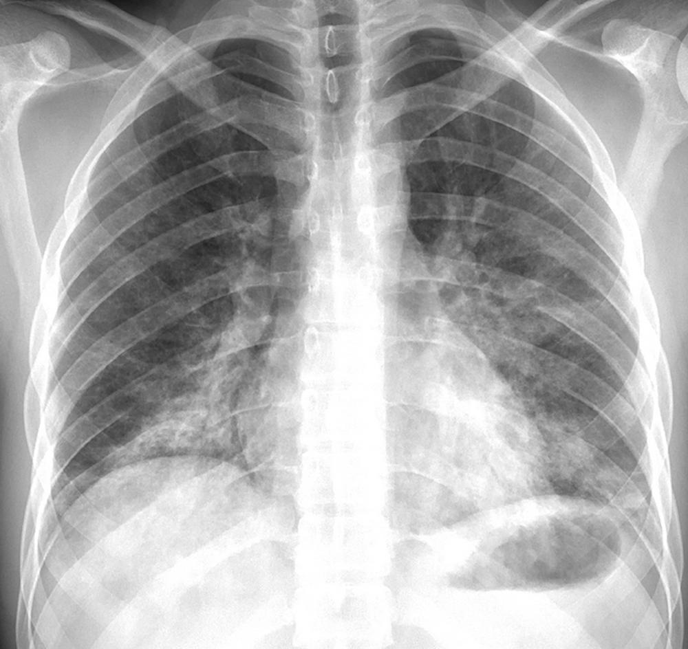
Figure 7 (from Radiology: Cardiothoracic Imaging). Posteroanterior chest radiograph in a 24-year-old man with a history of e-cigarette use shows bilateral reticular and predominantly ground-glass and airspace opacities distributed throughout both lower lungs and absence of pleural effusions.
High-res (TIF) version
(Right-click and Save As)
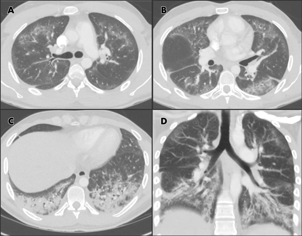
Figure 8 (from Radiology: Cardiothoracic Imaging). Chest CT angiographic images in a 24-year-old man with a history of e-cigarette use. A–C, Selected axial sections of the lung presented on lung windows reveal patchy ground-glass opacities distributed bilaterally with some, A, B, subpleural sparing and, C, consolidation in the lung bases. D, Coronal oblique thick-slab average intensity reconstruction shows the predominance of the findings in the basal lungs and demonstrates the peripheral subpleural sparing.
High-res (TIF) version
(Right-click and Save As)
Related Links: