3-D Printing Takes Imaging to the Next Dimension
At A Glance
- The use of 3-D printing in medicine is rapidly expanding.
- In the clinic, 3-D printing plays three major roles: surgical planning, patient-specific simulations and education.
- RSNA can offer peer-reviewed research, images, video and expert commentary to assist in developing news stories on 3-D printing in medicine.
- RSNA Media Relations
1-630-590-7762
media@rsna.org - Linda Brooks
1-630-590-7738
lbrooks@rsna.org
(May 1, 2017) — The use of 3-D printing in medicine is rapidly expanding. Applications of the technology in surgical preparation, patient-specific simulations and education are increasing exponentially. Experimental uses, such as the printing of body parts, are no longer the stuff of science fiction.
The Radiological Society of North America (RSNA) recognizes the value this technology brings to diagnosis and treatment of patients and has established a Special Interest Group to promote the highest quality medical 3-D printing applications via education and research, collaboration and research.
While the visual impact of 3-D printed models are captivating, the models offer far more than an aesthetic appeal. In the clinic, 3-D printing plays three major roles: surgical planning, patient-specific simulations and education.
In surgical planning, a model allows surgeons to understand a patient’s anatomy before operating. For instance, models can show how a tumor might be encasing critical nerves that allow an arm or leg to function, and 3-D printed cutting guides help take the guesswork out of a procedure. For patient-specific simulations, physicians can have two custom stents made based on a patient’s vascular anatomy and use one for a simulation. The additional planning can help reduce the significant expenses of operating room time.
“With 3-D printing, you take things that are unknowns before you go into the operating room and make them known,” said Jonathan M. Morris, M.D., assistant radiology professor at the Mayo Clinic in Rochester, Minn., and chair of the RSNA 3-D Printing Special Interest Group. “You decrease time in the operating room, decrease morbidity for the patient and realize huge cost savings and better patient outcomes.”
Medical teams use 3-D printed models to plan complex surgeries like the separation of conjoined twins and human face transplants. The technology is also being used to print living tissue, such as in trachea reconstructions.
Education is also a significant component of 3-D printing. Printed models can help a patient understand a procedure that can be difficult to describe in layperson’s terms. Pre-surgical study of models helps everyone on the team, from the scrub techs to the anesthesiologists. Models can even show pathologists where a tissue sample came from.
While researchers see 3-D printing playing a huge role in the future, it also provides us an astounding glimpse into the past, combining with CT technology to create more accurate recreations of dinosaur fossils.
Feel free to browse the selection of research and education articles on 3-D printing at the links below and contact the RSNA media relations team for interviews with leading experts in this exciting field.
Images and video from all of the research links are available for media use. Contact RSNA media relations for assistance.
3-D Printing Research
RSNA News: “3-D Scoliotic Spine Model Aids Pre-Surgical Planning in 8-Year-Old Girl”
RSNA News: "Defining Radiology's Role in the 3-D Printing Explosion"
RSNA Newsroom: "New Studies Provide More Insight into Zika Effects"
RSNA Newsroom: “Researchers Use 3-D Printing to Guide Human Face Transplants”
RSNA Newsroom: “Researchers Use CT and 3-D Printers to Recreate Dinosaur Fossils”
RSNA Newsroom: “CT and 3-D Printing Aid Surgical Separation of Conjoined Twins”
RSNA News: “3-D Printed Models Move Complex Surgery in Exciting New Directions”
RSNA News: “3-D Printed Models Add Colorful New Dimension to Complex Surgery”
RSNA News: “3-D MRI Fetal Images Superior in Quality to 3-D US”
RSNA Newsroom: 3-D-Printed Prosthetic Implants Could Improve Treatment for Hearing Loss
3-D Printing Background
Radiographics: “Medical 3-D Printing for the Radiologist”
Radiographics: “Three-dimensional Physical Modeling: Applications and Experience at Mayo Clinic”
Radiographics: “Measuring and Establishing the Accuracy and Reproducibility of 3D Printed Medical Models”
RSNA is an association of over 54,600 radiologists, radiation oncologists, medical physicists and related scientists, promoting excellence in patient care and health care delivery through education, research and technologic innovation. The Society is based in Oak Brook, Ill. (RSNA.org)
Resources:
Images (.JPG and .TIF format)
and Video clips (.mp4 format)

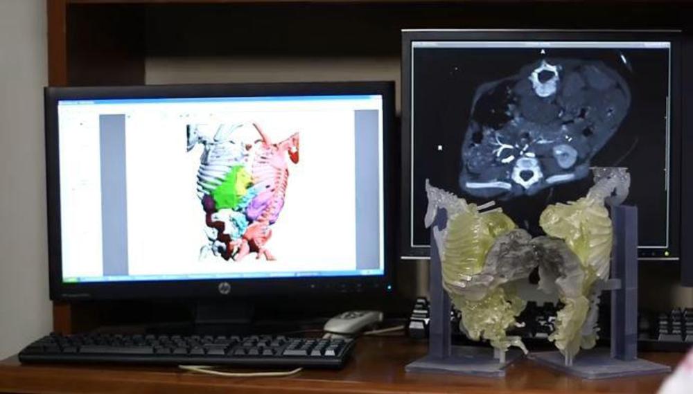
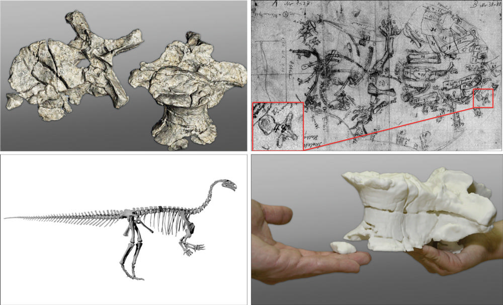
Figure 3. (Clockwise) 1) Virtual 3-D reconstructions of fossilized vertebral body after surrounding sediment matrix and protective plaster have been digitally removed through segmentation of CT dataset. Multiple fracture lines and dislocated fragment from anterior rim of vertebral body are clearly visible. 2) Digitized image of original excavation field map from Halberstadt, 1923. Inset is magnification of fossilized vertebra. 3) Illustration of Plateosaurus skeleton. 4) 3-D print of vertebral body.
High-res (TIF) version
(Right-click and Save As)
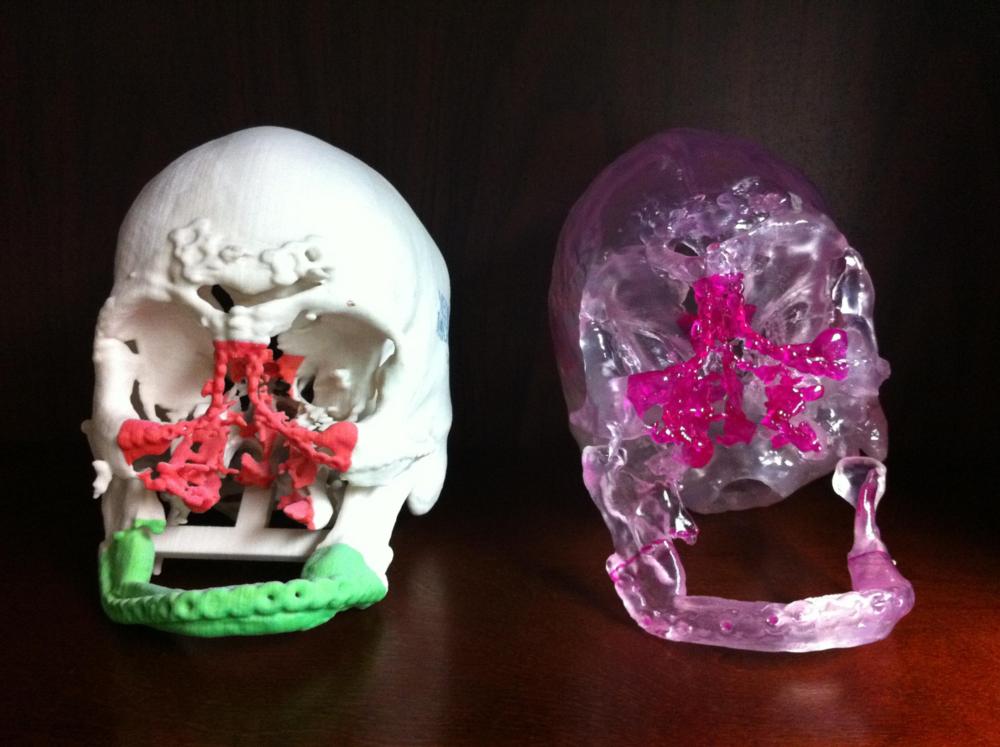
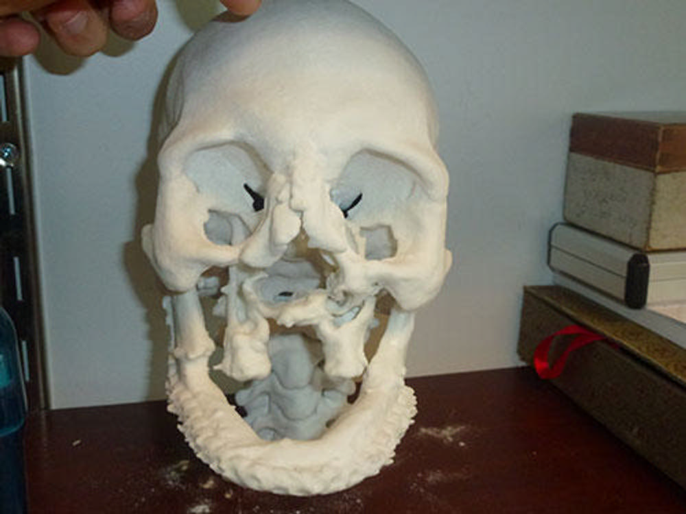
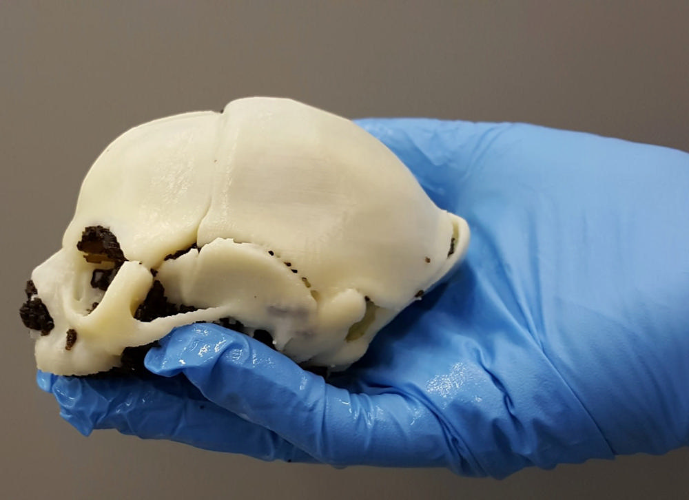
Figure 6. 3-D print of post-natal CT of infant with microcephaly due to the Zika virus.
High-res (TIF) version
(Right-click and Save As)
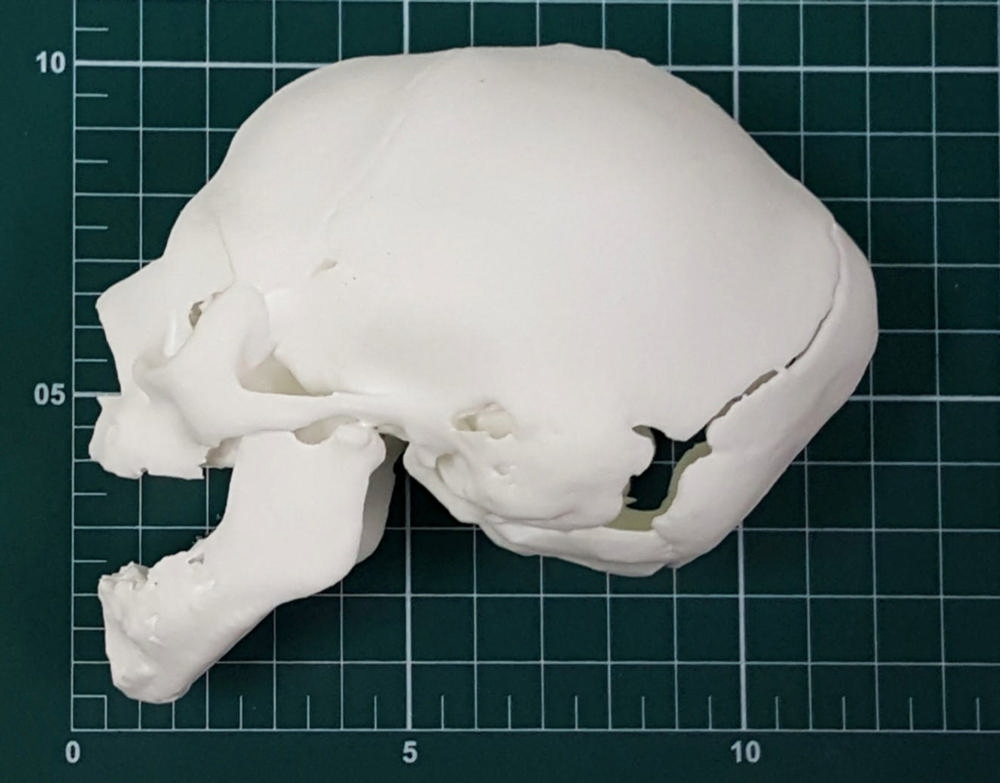
Figure 7. 3-D print of post-natal CT of infant with microcephaly due to the Zika virus.
High-res (TIF) version
(Right-click and Save As)
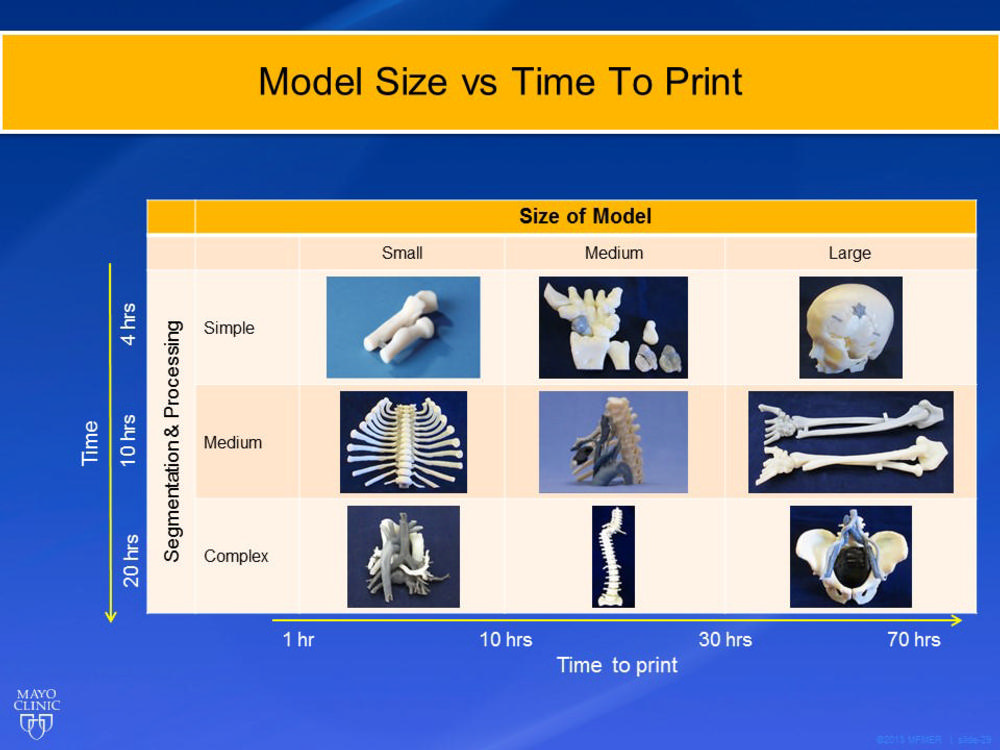
Figure 8. Model size versus time to print. Created by Mayo Clinic. Copyright 2017 Mayo Foundation for Medical Education and Research. Used with permission.
High-res (TIF) version
(Right-click and Save As)
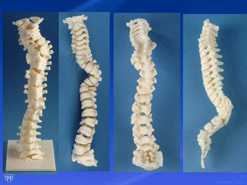
Figure 9. 3-D print of spine used for orthopedic surgery. Created by Mayo Clinic. Copyright 2017 Mayo Foundation for Medical Education and Research. Used with permission.
High-res (TIF) version
(Right-click and Save As)
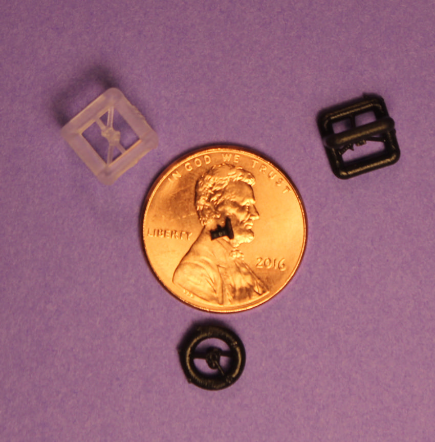
Figure 10. Size comparison between 3-D printed prosthesis ear implant and a penny.
High-res (TIF) version
(Right-click and Save As)
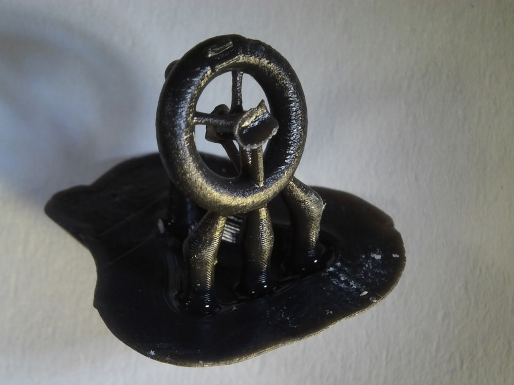
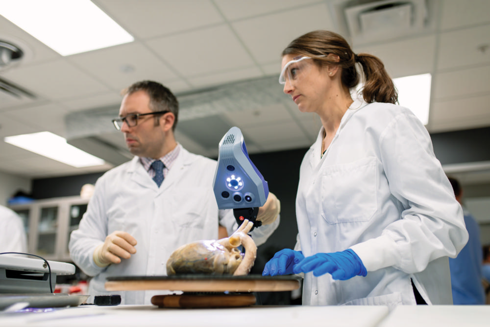
Figure 12. Pathologist Joseph Maleszewski, M.D., Ph.D., associate professor at Mayo Clinic School of Graduate Medical Education, scans a heart for 3-D reconstruction. Dr. Maleszewski is pictured with Melanie C. Bois, M.D., a cardiovascular fellow. Image courtesy of the Mayo Clinic.
High-res (TIF) version
(Right-click and Save As)
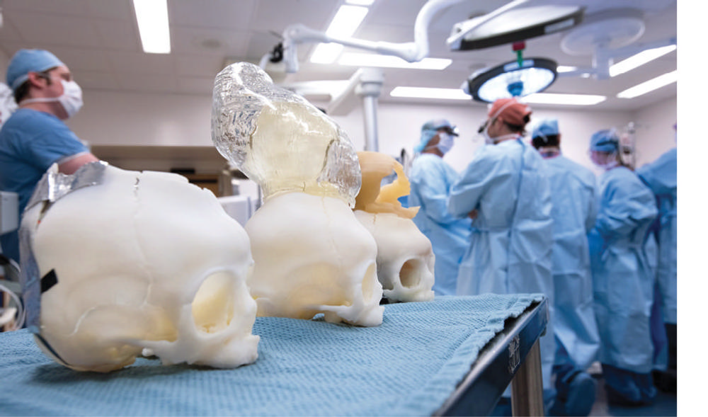
Figure 13. The surgical teams at Boston Children's Hospital use 3-D models to practice on rarely seen conditions that even experienced surgeons likely have not faced, including craniosynostosis, a condition in which the bones of the skull fuse in an abnormal fashion. Image courtesy of Boston Children's Hospital.
High-res (TIF) version
(Right-click and Save As)
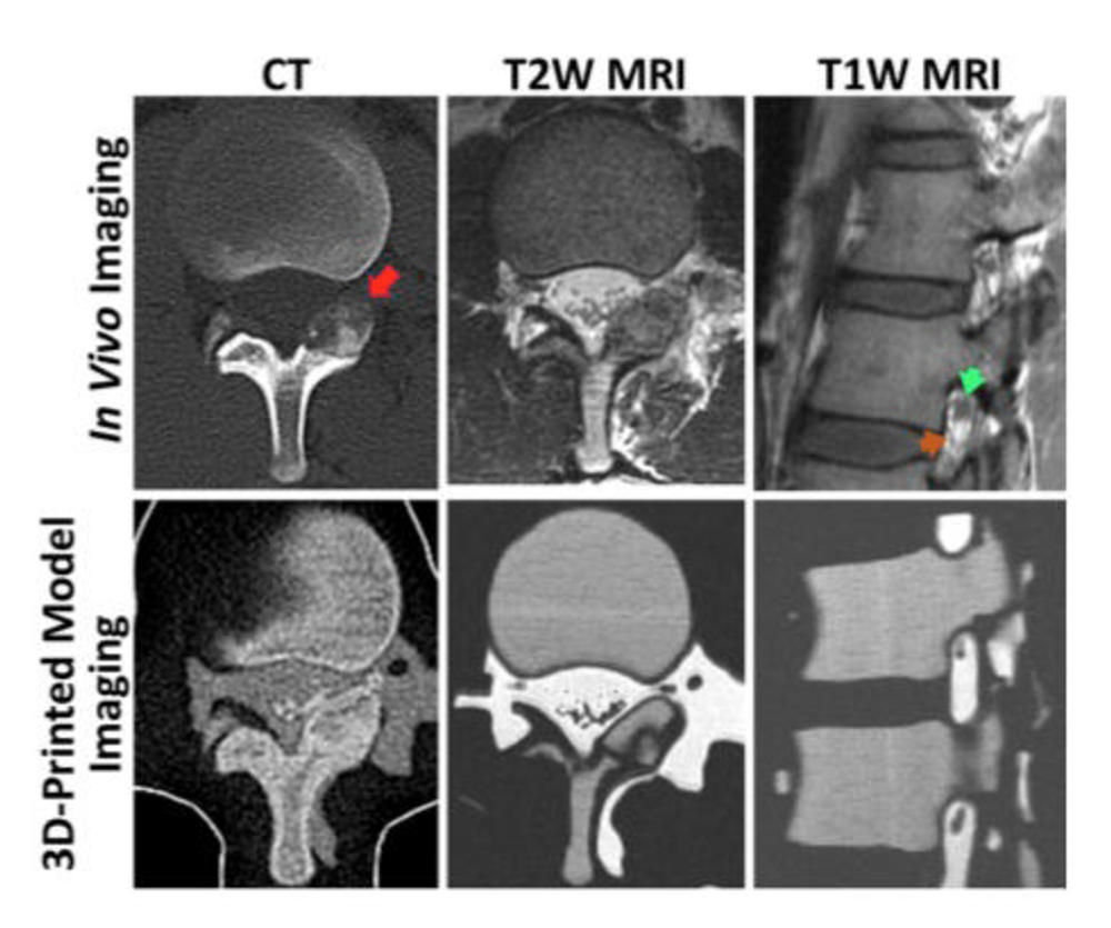
Figure 14. The 3-D printing process at Brigham and Women's Hospital: Top row, from left: CT and MRI of a patient with L1 left lamina osteoblastoma (red arrow). Segmenting the individual tissues in these images and using 3-D printing to see the images using different radiopaque materials, some MR-visible and some not, produces a printed model (bottom) that replicates the signal densities/intensities of the tissues similar to the in vivo images, including the tumor, adipose tissue including foraminal fat (brown arrows) and spinal nerves (green arrows). Image courtesy of Brigham and Women's Hospital.
High-res (TIF) version
(Right-click and Save As)
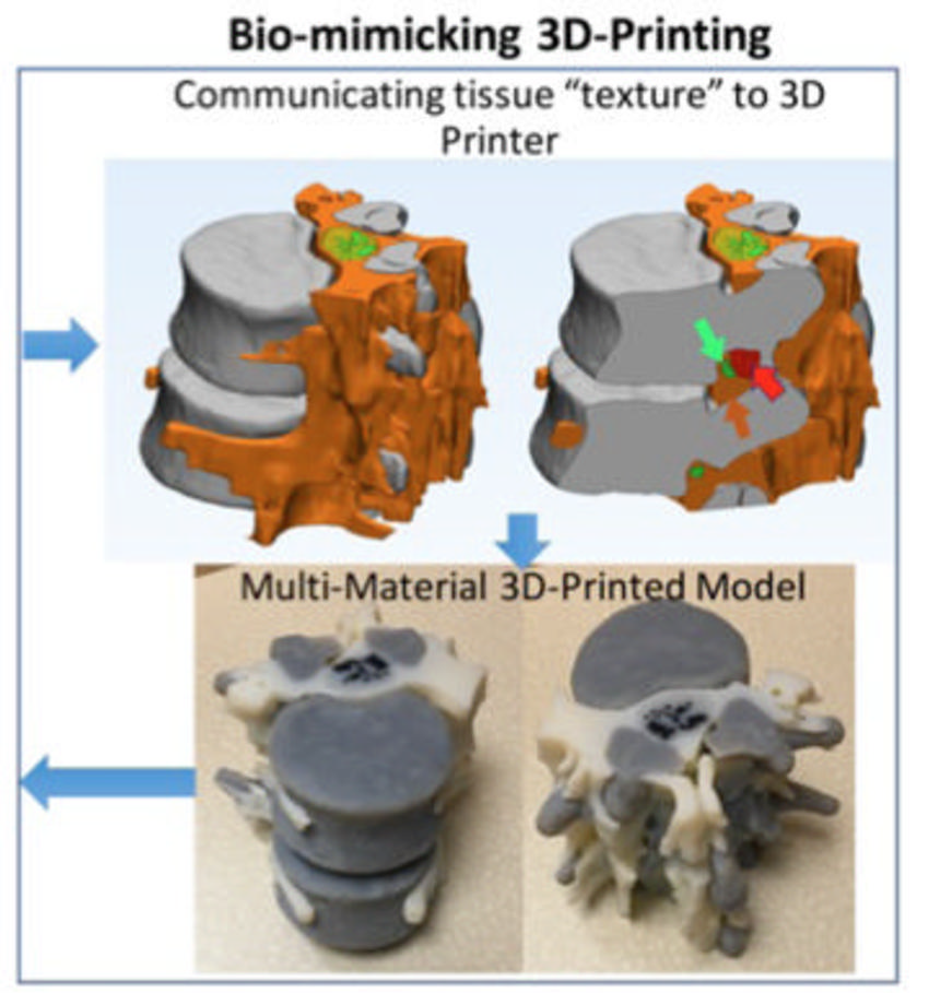
Figure 15. 3-D printed model of a patient with L1 left lamina osteoblastoma (red arrow). The model replicates the signal densities/intensities of the tissues similar to the in vivo images, including the tumor, adipose tissue including foraminal fat (brown arrows) and spinal nerves (green arrows). The 3-D printed model was used to simulate the CT-guided power drilling and subsequent MRI–monitored cryoablation to ensure that a tumor could be ablated without the ice ball impinging on the nerve root (green arrow). Image courtesy of Brigham and Women's Hospital.
High-res (TIF) version
(Right-click and Save As)
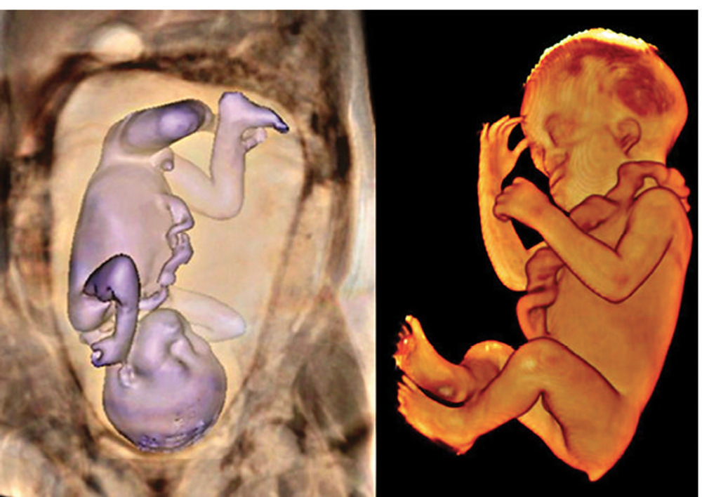
Figure 16. 3-D MRI showing a 27-week fetus with brachycephaly, low ear implantation, syndromic profile and vestigial tail.
High-res (TIF) version
(Right-click and Save As)
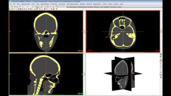
Video 1. CT Reconstruction for 3D Printing. Video provided by Materialise and Mimics Software.
Download.mp4
(Right-click and Save As)
Video 2. 3D Model of Mata Conjoined Twins. Used with the permission of Texas Children’s Hospital.
Download.mp4
(Right-click and Save As)
Video 3. Dr. Frank Rybicki explains other medical and surgical uses for 3-D models.
Download.mp4
(Right-click and Save As)