RSNA Press Release
- CT scans can be used with 3-D printers to make accurate copies of fossilized bones.
- The CT study provided valuable information about the condition and integrity of the fossil.
- The CT dataset helped the researchers build an accurate reconstruction of the fossil with selective laser sintering, a technology that uses a high-powered laser to fuse together materials to make a 3-D object.
Researchers Use CT and 3-D Printers to Recreate Dinosaur Fossils
Released: November 20, 2013
| Media Contacts: | |
| RSNA Media Relations: | 1-630-590-7762 |
| Linda Brooks 1-630-590-7738 lbrooks@rsna.org |
Maureen Morley 1-630-590-7754 mmorley@rsna.org |
OAK BROOK, Ill. — Data from computed tomography (CT) scans can be used with three-dimensional (3-D) printers to make accurate copies of fossilized bones, according to new research published online in the journal Radiology.
Fossils are often stored in plaster casts, or jackets, to protect them from damage. Getting information about a fossil typically requires the removal of the plaster and all the sediment surrounding it, which can lead to loss of material or even destruction of the fossil itself.
German researchers studied the feasibility of using CT and 3-D printers to nondestructively separate fossilized bone from its surrounding sediment matrix and produce a 3-D print of the fossilized bone itself.
"The most important benefit of this method is that it is non-destructive, and the risk of harming the fossil is minimal," said study author Ahi Sema Issever, M.D., from the Department of Radiology at Charité Campus Mitte in Berlin. "Also, it is not as time-consuming as conventional preparation."
Dr. Issever and colleagues applied the method to an unidentified fossil from the Museum für Naturkunde, a major natural history museum in Berlin. The fossil and others like it were buried under rubble in the basement of the museum after a World War II bombing raid. Since then, museum staff members have had difficulty sorting and identifying some of the plaster jackets.
Researchers performed CT on the unidentified fossil with a 320-slice multi-detector system. The different attenuation, or absorption of radiation, through the bone compared with the surrounding matrix enabled clear depiction of a fossilized vertebral body.
After studying the CT scan and comparing it to old excavation drawings, the researchers were able to trace the fossil's origin to the Halberstadt excavation, a major dig from 1910 to 1927 in a clay pit south of Halberstadt, Germany. In addition, the CT study provided valuable information about the condition and integrity of the fossil, showing multiple fractures and destruction of the front rim of the vertebral body.
Furthermore, the CT dataset helped the researchers build an accurate reconstruction of the fossil with selective laser sintering, a technology that uses a high-powered laser to fuse together materials to make a 3-D object.
Dr. Issever noted that the findings come at a time when advances in technology and cheaper availability of 3-D printers are making them more common as a tool for research. Digital models of the objects can be transferred rapidly among researchers, and endless numbers of exact copies may be produced and distributed, greatly advancing scientific exchange, Dr. Issever said. The technology also potentially enables a global interchange of unique fossils with museums, schools and other settings.
"The digital dataset and, ultimately, reproductions of the 3-D print may easily be shared, and other research facilities could thus gain valuable informational access to rare fossils, which otherwise would have been restricted," Dr. Issever said. "Just like Gutenberg's printing press opened the world of books to the public, digital datasets and 3-D prints of fossils may now be distributed more broadly, while protecting the original intact fossil."
# # #
"Reviving the Dinosaur: Virtual Reconstruction and Three-dimensional Printing of a Dinosaur Vertebra." Collaborating with Dr. Issever were René Schilling, M.D., Benjamin Jastram, Dipl.-lng., Oliver Wings, Dr. rer. nat., and Daniela Schwarz, Dr. rer. nat.
Radiology is edited by Herbert Y. Kressel, M.D., Harvard Medical School, Boston, Mass., and owned and published by the Radiological Society of North America, Inc. (http://radiology.rsna.org/)
RSNA is an association of more than 53,000 radiologists, radiation oncologists, medical physicists and related scientists, promoting excellence in patient care and health care delivery through education, research and technologic innovation. The Society is based in Oak Brook, Ill. (RSNA.org)
For patient-friendly information on CT, visit RadiologyInfo.org.
 Figure 1. Illustration of Plateosaurus skeleton. High-res (TIF) version (Right-click and Save As) |
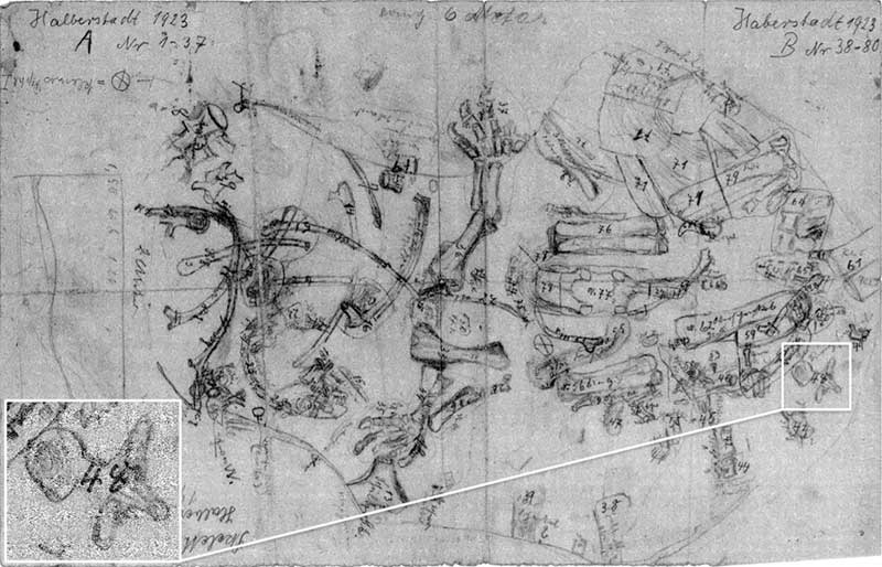 Figure 2. Digitized image of original excavation field map from Halberstadt, 1923. Inset is magnification of fossilized vertebra. High-res (TIF) version (Right-click and Save As) |
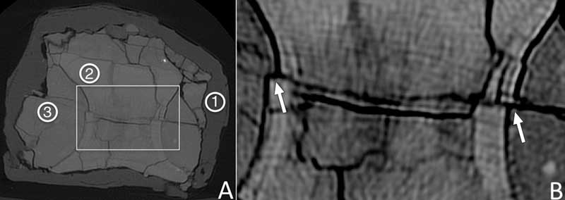 Figure 3. Axial CT scans of fossil. A, CT scan (window level, 4698 HU; window width, 13 493 HU) covers full scale of attenuation and depicts entire inner structure of scanned object, including surrounding plaster jacket, sediment matrix, and fossilized vertebral body. To obtain representative attenuation measurements, circular ROIs were placed in surrounding protective plaster (1), body of vertebra (2), and surrounding fossil¬ized sediment matrix (3). B, To increase contrast between fossilized vertebral body and fossilized surrounding sediment matrix, a section—square in A—is depicted with window level of 2600 HU and window width of 2600 HU. Arrows in B indicate displacement of ventral and dorsal halves of vertebra (see arrows in Fig 6, F). High-res (TIF) version (Right-click and Save As) |
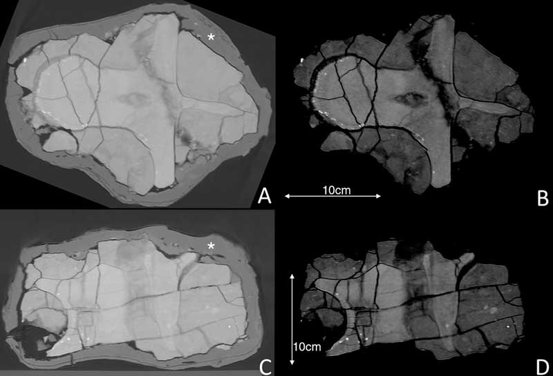 Figure 4. A, B, Coronal and, C, D, sagittal reconstructions of fossil with different window levels and widths. In A and C, window levels and widths were set to a full dynamic range (A: window level = 1370 HU, window width = 6836 HU; C: window level = 1113 HU, window width = 6322 HU). In B and D, the window level and width were set to 2600 HU. * indicates surrounding hard plaster. Numerous fracture lines passing through fossil are clearly visible. High-res (TIF) version (Right-click and Save As) |
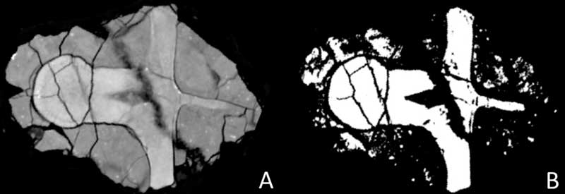 Figure 5. Coronal views of fossil. A, To segment the dataset for the 3D print and to improve visual differentiation, attenuation values were first remapped into a smaller range. B, Recalibration of brightness and contrast values further improves differentiation between fossilized vertebral body and its surrounding sediment matrix. High-res (TIF) version (Right-click and Save As) |
 Figure 6. A, C, E, Enlarged sections of squares in B, D, and F, respectively. In coronal views (A and C), fracture lines (arrows) pass through surrounding plaster jacket (*) and extend into fossil. Arrows on sagittal view (F) mark major fracture zone that goes through entire fossil, causing displacement of ventral and dorsal halves as marked by arrows in Figure 3, B. In enlarged section of image (E), a void between the surrounding plaster (*) and the underlying—apparently destroyed—anterior part of fossil is clearly visible. Multiple small fragments can be identified within the void. High-res (TIF) version (Right-click and Save As) |
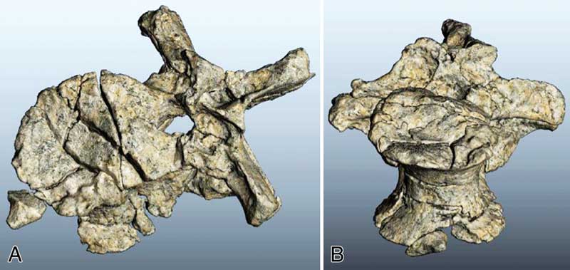 Figure 7. Virtual 3D reconstructions of fossilized vertebral body after surrounding sediment matrix and protective plaster have been digitally removed through segmentation of CT dataset. Multiple fracture lines and dislocated fragment from anterior rim of vertebral body are clearly visible. High-res (TIF) version (Right-click and Save As) |
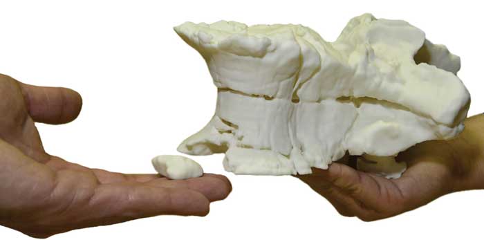 Figure 8. 3D print of vertebral body. High-res (TIF) version (Right-click and Save As) |
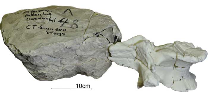 Figure 9. 3D print next to the original unprepared and erroneously labeled plaster jacket. High-res (TIF) version (Right-click and Save As) |
 PDF
PDF