RSNA Press Release
- Research suggests that migraines are related to brain abnormalities present at birth and others that develop over time.
- Migraine patients in the study showed reduced cortical thickness and surface area in regions related to pain processing.
- More than 300 million people suffer from migraines worldwide.
MRI Shows Brain Abnormalities in Migraine Patients
Released: March 26, 2013
| Media Contacts: | |
| RSNA Media Relations: | 1-630-590-7762 |
| Linda Brooks 1-630-590-7738 lbrooks@rsna.org |
Maureen Morley 1-630-590-7754 mmorley@rsna.org |
OAK BROOK, Ill. — A new study suggests that migraines are related to brain abnormalities present at birth and others that develop over time. The research is published online in the journal Radiology.
Migraines are intense, throbbing headaches, sometimes accompanied by nausea, vomiting and sensitivity to light. Some patients experience auras, a change in visual or sensory function that precedes or occurs during the migraine. More than 300 million people suffer from migraines worldwide, according to the World Health Organization.

Previous research on migraine patients has shown atrophy of cortical regions in the brain related to pain processing, possibly due to chronic stimulation of those areas. Cortical refers to the cortex, or outer layer of the brain.
Much of that research has relied on voxel-based morphometry, which provides estimates of the brain's cortical volume. In the new study, Italian researchers used a different approach: a surface-based MRI method to measure cortical thickness.
"For the first time, we assessed cortical thickness and surface area abnormalities in patients with migraine, which are two components of cortical volume that provide different and complementary pieces of information," said Massimo Filippi, M.D., director of the Neuroimaging Research Unit at the University Ospedale San Raffaele and professor of neurology at the University Vita-Salute's San Raffaele Scientific Institute in Milan. "Indeed, cortical surface area increases dramatically during late fetal development as a consequence of cortical folding, while cortical thickness changes dynamically throughout the entire life span as a consequence of development and disease."
Dr. Filippi and colleagues used magnetic resonance imaging (MRI) to acquire T2-weighted and 3-D T1-weighted brain images from 63 migraine patients and 18 healthy controls. Using special software and statistical analysis, they estimated cortical thickness and surface area and correlated it with the patients' clinical and radiologic characteristics.
Compared to controls, migraine patients showed reduced cortical thickness and surface area in regions related to pain processing. There was only minimal anatomical overlap of cortical thickness and cortical surface area abnormalities, with cortical surface area abnormalities being more pronounced and distributed than cortical thickness abnormalities. The presence of aura and white matter hyperintensities—areas of high intensity on MRI that appear to be more common in people with migraine—was related to the regional distribution of cortical thickness and surface area abnormalities, but not to disease duration and attack frequency.
"The most important finding of our study was that cortical abnormalities that occur in patients with migraine are a result of the balance between an intrinsic predisposition, as suggested by cortical surface area modification, and disease-related processes, as indicated by cortical thickness abnormalities," Dr. Filippi said. "Accurate measurements of cortical abnormalities could help characterize migraine patients better and improve understanding of the pathophysiological processes underlying the condition."
Additional research is needed to fully understand the meaning of cortical abnormalities in the pain processing areas of migraine patients, according to Dr. Filippi.
"Whether the abnormalities are a consequence of the repetition of migraine attacks or represent an anatomical signature that predisposes to the development of the disease is still debated," he said. "In my opinion, they might contribute to make migraine patients more susceptible to pain and to an abnormal processing of painful conditions and stimuli."
The researchers are conducting a longitudinal study of the patient group to see if their cortical abnormalities are stable or tend to worsen over the course of the disease. They are also studying the effects of treatments on the observed modifications of cortical folding and looking at pediatric patients with migraine to assess whether the abnormalities represent a biomarker of the disease.
# # #
"Cortical Abnormalities in Patients with Migraine: A Surface-based Analysis." Collaborating with Dr. Filippi were Roberta Messina, M.D., Maria A. Rocca, M.D., Bruno Colombo, M.D., Paola Valsasina, M.Sc., Mark A. Horsfield, Ph.D., Massimilano Copetti, Ph.D., Andrea Falini, M.D., and Giancarlo Comi, M.D.
Radiology is edited by Herbert Y. Kressel, M.D., Harvard Medical School, Boston, Mass., and owned and published by the Radiological Society of North America, Inc. (http://radiology.rsna.org/)
RSNA is an association of more than 51,000 radiologists, radiation oncologists, medical physicists and related scientists promoting excellence in patient care and health care delivery through education, research and technologic innovation. The Society is based in Oak Brook, Ill. (RSNA.org)
For patient-friendly information on MRI, visit RadiologyInfo.org.
Images
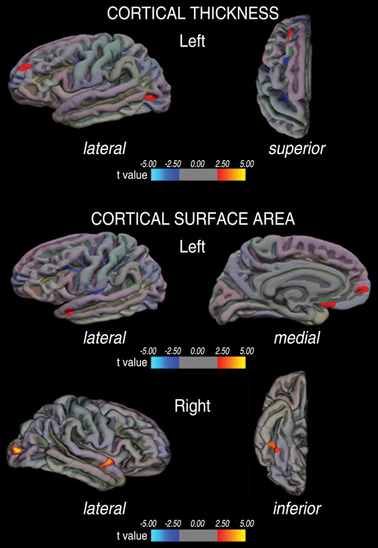 Figure 1 Vertex-by-vertex analysis shows regional differences in cortical thickness and cortical surface area in patients with migraine compared with healthy control subjects (P < .01) represented on an averaged brain map. Regions of increased cortical thickness or surface area are shown in red (color coded according to t value), and regions of decreased cortical thickness or surface area are shown in blue (color-coded according to t value). Only the most representative views are shown. High-res (TIF) version (Right-click and Save As) |
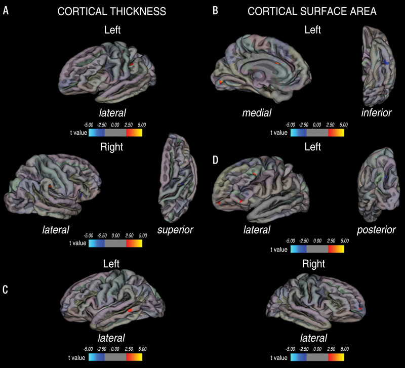 Figure 2 Vertex-by-vertex analysis shows regional differences in, A, C, cortical thickness and, B, D, cortical surface area between patients with migraine with aura, patients with migraine without aura, and healthy control subjects represented on an averaged brain map. Regions of increased cortical thickness or surface area are shown in red (color coded according to t value), and regions of decreased cortical thickness or surface area are shown in blue (color coded according to t value). A, B, Data in patients with aura versus data in both healthy control subjects and patients without aura. C, D, Data in patients without aura versus data in both healthy control subjects and patients with aura. Only the most representative views are shown. High-res (TIF) version (Right-click and Save As) |
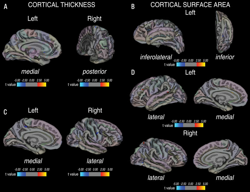 Figure 3 Vertex-by-vertex analysis shows regional differences in, A, C, cortical thickness and, B, D, cortical surface area between patients with migraine with WMHs, patients with migraine without WMHs, and healthy control subjects represented on an averaged brain map. Regions of increased cortical thickness or surface area are shown in red (color coded according to t value), and regions of decreased cortical thickness or surface area are shown in blue (color coded according to t value). A, B, Data in patients with migraine with WMHs versus data in both healthy control subjects and patients with migraine without WMHs. C, D, Data in patients with migraine without WMHs versus data in both healthy control subjects and patients with migraine with WMHs. Only the most representative views are shown. High-res (TIF) version (Right-click and Save As) |
 PDF
PDF