RSNA Press Release
- Too much physical activity may damage knee cartilage over time, but too little activity may also accelerate degeneration.
- Along with the findings on changes in knee cartilage, the study also highlighted the potential of MRI-based T2 relaxation times as an early indicator of cartilage degeneration.
- T2 relaxation times can potentially detect cartilage changes at an earlier stage when still reversible.
Too Much or Too Little Activity Bad for Knees
Released: November 26, 2012
| Media Contacts: | RSNA Newsroom | 1-312-949-3233 |
| Before 11/24/12 or after 11/29/12: | RSNA Media Relations: | 1-630-590-7762 |
| |
Linda Brooks 1-630-590-7738 lbrooks@rsna.org |
Maureen Morley 1-630-590-7754 mmorley@rsna.org |
CHICAGO—Both very high and very low levels of physical activity can accelerate the degeneration of knee cartilage in middle-aged adults, according to a new study presented at the annual meeting of the Radiological Society of North America (RSNA).


Nearly one in every two people in the U.S. may develop knee osteoarthritis by age 85, according to the Centers for Disease Control and Prevention. By 2030, an estimated 67 million Americans over the age of 18 are projected to have physician-diagnosed arthritis.
Researchers at the University of California in San Francisco (UCSF) previously had found an association between physical activity and cartilage degeneration. But that study focused on one point in time.
For the new study, the UCSF researchers looked at changes in knee cartilage among a group of middle-aged adults over a four-year period. They used magnetic resonance imaging (MRI)-based T2 relaxation times to track the evolution of early degenerative cartilage changes in the knee.
"T2 relaxation times generated from MR images allow for analysis of the biochemical and molecular composition of cartilage," said Wilson Lin, B.S., research fellow and medical student at UCSF. "There is increased water mobility in damaged cartilage, and increased water mobility results in increased T2 relaxation time."
The researchers analyzed 205 patients, age 45 to 60, from the UCSF-based Osteoarthritis Initiative, a nationwide study funded by the National Institutes of Health on the prevention and treatment of knee osteoarthritis. Participants used a questionnaire to record their physical activity. The researchers measured T2 values of cartilage at the patella, femur and tibia of the right knee joint at baseline and at two- and four-year visits.
According to the results of the study, participating frequently in high-impact activities, such as running, appears associated with more degenerated cartilage and potentially a higher risk for development of osteoarthritis.
"When we compared the scores among groups, we found an accelerated progression of T2 relaxation times in those who were the most physically active," said Thomas M. Link, M.D., professor of radiology and chief of musculoskeletal imaging at UCSF. "Those who had very low levels of activity also had accelerated progression of T2 values. This suggests that there may be an optimal level of physical activity to preserve the cartilage."
The results open up numerous areas for future inquiry, including analysis of the impact of specific types of physical activity on knee cartilage health. For instance, some of the participants in the Osteoarthritis Initiative wore an accelerometer, a device with a motion sensor to record physical activity.
"In this study, we used the subjective measure of a questionnaire," Lin said. "The accelerometers provide a more objective way to measure physical activity."
Along with the findings on changes in knee cartilage, the study also highlighted the potential of T2 relaxation times as an early indicator of cartilage degeneration.
"Standard MRI shows cartilage defects that are irreversible," Dr. Link said. "The exciting thing about the new cartilage T2 measurements is that they give us information on a biochemical level, thus potentially detecting changes at an earlier stage when they may still be reversible."
Dr. Link noted that people who have a higher risk for osteoarthritis (such as family history of total joint replacement, obesity, history of knee injury or surgery) can reduce their risk for cartilage degeneration by maintaining a healthy weight and avoiding risky activities and strenuous, high-impact exercise.
"Lower impact sports, such as walking or swimming, are likely more beneficial than higher impact sports, such as running or tennis, in individuals at risk for osteoarthritis," he said.
Coauthors are Waraporn Srikhum, M.D., Charles E. McCulloch, Ph.D., Michael Neitt, Ph.D., John Lynch, Ph.D., Gabby B. Joseph, Ph.D., and Hamza Alizai, M.D.
# # #
Note: Copies of RSNA 2012 news releases and electronic images will be available online at RSNA.org/press12 beginning Monday, Nov. 26.
RSNA is an association of more than 50,000 radiologists, radiation oncologists, medical physicists and related scientists promoting excellence in patient care and health care delivery through education, research and technologic innovation. The Society is based in Oak Brook, Ill. (RSNA.org)
Editor's note: The data in these releases may differ from those in the published abstract and those actually presented at the meeting, as researchers continue to update their data right up until the meeting. To ensure you are using the most up-to-date information, please call the RSNA Newsroom at 1-312-949-3233.
For patient-friendly information on MRI of the knee, visit RadiologyInfo.org.
| Abstract: |
Video clips
- (.mp4 format)
- Video clip (28,562 Kbyte)
Dr. Thomas Link explains how too much or too intense physical activity can be detrimental for knees.
Images (.JPG format)
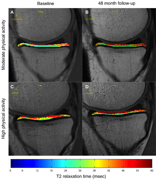 Figure 1. All four diagrams show T2 color maps of knee cartilage located at the medial tibia. An individual from the high physical activity group (C and D) shows a greater increase in T2 relaxation time compared to an individual from the moderate physical activity group (A and B). High-res (TIF) version (Right-click and Save As) |
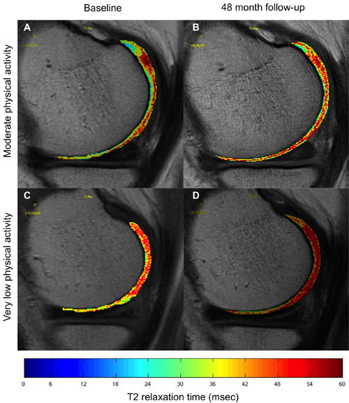 Figure 1. All four diagrams show T2 color maps of knee cartilage located at the medial femoral condyle. An individual from the very low physical activity group (C and D) shows a greater increase in T2 relaxation time compared to an individual from the moderate physical activity group (A and B). High-res (TIF) version (Right-click and Save As) |
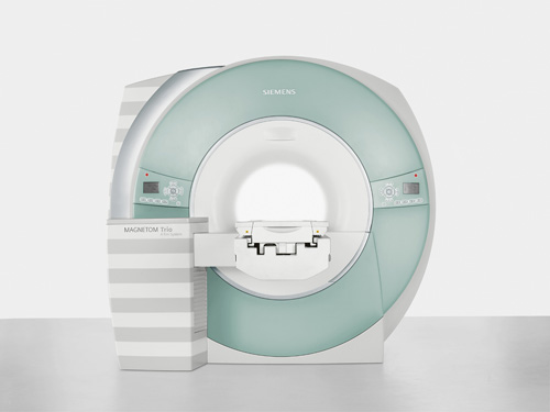 Figure 3. Photograph of a Siemens Megnetom Trio MRI Scanner. High-res (TIF) version (Right-click and Save As) |
 PDF
PDF