RSNA Press Release
- MR spectroscopy may allow the diagnosis of chronic traumatic encephalopathy (CTE) before death.
- CTE is a degenerative brain disorder caused by repeated brain trauma, including concussions and multiple blows to the head, such as those found in contact sports.
- Approximately 3.8 million sports- and recreation-related concussions occur in the U.S. each year, and 22 professional football players have been diagnosed with CTE.
Virtual Biopsy May Allow Earlier Diagnosis of Brain Disorder in Athletes
Released: December 1, 2010
| Media Contacts: | RSNA Newsroom | 1-312-949-3233 |
| Before 11/27/2010 or after 12/02/2010: | RSNA Media Relations: | 1-630- 590-7762 |
| |
Linda Brooks 1-630-590-7738 lbrooks@rsna.org |
Maureen Morley 1-630-590-7754 mmorley@rsna.org |
CHICAGO — In a study of ex-pro athletes, researchers found that a specialized imaging technique called magnetic resonance spectroscopy (MRS) may help diagnose chronic traumatic encephalopathy (CTE), a disorder caused by repetitive head trauma that currently can only be definitively diagnosed at autopsy. Results of the study were presented today at the annual meeting of the Radiological Society of North America (RSNA).
"The devastating effects of brain injuries suffered by pro football players who repeatedly suffered concussions and subconcussive brain trauma during their careers have put the spotlight on CTE," said Alexander P. Lin, Ph.D., a principal investigator at the Center for Clinical Spectroscopy at Brigham and Women's Hospital in Boston. "However, blows to the head suffered by all athletes involved in contact sports are of increasing concern."
According to the Centers for Disease Control and Prevention, an estimated 3.8 million sports- and recreation-related concussions occur in the U.S. each year. In addition, subclinical concussions—injuries that cannot be diagnosed as concussions but have similar effects—are often unrecognized.
Studies have shown that individuals who suffer repetitive brain trauma are more likely to experience ongoing problems, from permanent brain damage to long-term disability.
CTE is a degenerative brain disease caused by repeated brain trauma and marked by a buildup of abnormal proteins in the brain. CTE has been associated with memory difficulty, impulsive and erratic behavior, depression and eventually, dementia.
"Cumulative head trauma invokes changes in the brain, which over time can result in a progressive decline in memory and executive functioning in some individuals," Dr. Lin said. "MRS may provide us with noninvasive, early detection of CTE before further damage occurs, thus allowing for early intervention."
In Dr. Lin's study, conducted in collaboration with the Boston University Center for the Study of Traumatic Encephalopathy (CSTE), five retired professional male athletes from football, wrestling and boxing with suspected CTE and five age- and size-matched controls between the ages of 32 and 55 were examined with MRS. In MRS, sometimes referred to as "virtual biopsy," a powerful magnetic field and radio waves are used to extract information about chemical compounds within the body, using a clinical MR scanner.
The results revealed that compared with the brains of the control patients, the brains of the former athletes with suspected CTE had increased levels of choline, a cell membrane nutrient that signals the presence of damaged tissue, and glutamate/glutamine, or Glx. MRS also revealed altered levels of gamma-aminobutyric acid (GABA), aspartate, and glutamate in the brains of former athletes.
"By helping us identify the neurochemicals that may play a role in CTE, this study has contributed to our understanding of the pathophysiology of the disorder," Dr. Lin said.
For example, the amino acid and neurotransmitter glutamate is involved in most aspects of normal brain function and must be present in the right places and at the right concentration in order for the brain to be healthy — too much or too little can be harmful.
"Being able to diagnose CTE could help athletes of all ages and levels, as well as war veterans who suffer mild brain injuries, many of which go undetected," Dr. Lin said.
Results of CSTE neuropathological studies of retired football players and other athletes have led to significant changes in the NFL, as well as collegiate and youth sports. Recently, the researchers found evidence of CTE in 21-year-old Owen Thomas, the University of Pennsylvania football captain who committed suicide in April 2010.
Coauthors are Saadallah Ramadan, Ph.D., Hayden Box, B.S., Peter Stanwell, Ph.D., and Robert Stern, Ph.D. Dr. Lin's research team is led by Carolyn Mountford, D.Phil. Other collaborators include Ann McKee, M.D., Robert Cantu, M.D., and Christopher Nowinski.
# # #
Note: Copies of RSNA 2010 news releases and electronic images will be available online at RSNA.org/press10 beginning Monday, Nov. 29.
RSNA is an association of more than 44,000 radiologists, radiation oncologists, medical physicists and related scientists committed to excellence in patient care through education and research. The Society is based in Oak Brook, Ill. (RSNA.org)
Editor's note: The data in these releases may differ from those in the printed abstract and those actually presented at the meeting, as researchers continue to update their data right up until the meeting. To ensure you are using the most up-to-date information, please call the RSNA Newsroom at 1-312-949-3233.
For patient-friendly information on MRI and MRS, visit RadiologyInfo.org.
| Abstract: |
Video clips
- Video clip (1.71 Mbyte)
Dr. Alexander Lin explains chronic traumatic encephalopathy (CTE). - Video clip (0.84 Mbyte)
Dr. Alexander Lin describes the symptoms of chronic traumatic encephalopathy (CTE). - Video clip (1.35 Mbyte)
Dr. Alexander Lin explains the procedure for diagnosing chronic traumatic encephalopathy (CTE). - Video clip (1.38 Mbyte)
Dr. Alexander Lin discusses the significance of the study. - Video clip (1.9 Mbyte)
Dr. Alexander Lin explains what happens during MR spectroscopy. - Video clip (0.97 Mbyte)
Dr. Alexander Lin shares the results of the study. - Video clip (1.18 Mbyte)
Former NFL player Brent Boyd describes his football career path. - Video clip (2.76 Mbyte)
Former NFL player Brent Boyd discusses experiencing his first concussion during a football game. - Video clip (2.35 Mbyte)
Former NFL player Brent Boyd describes how his first concussion affected him. - Video clip (3.82 Mbyte)
Former NFL player Brent Boyd discusses the impact of his injuries on his quality of life. - Video clip (1.25 Mbyte)
Former NFL player Brent Boyd shares his concern for youth involved in contact sports. - Video clip (0.99 Mbyte)
Former NFL player Brent Boyd shares his advice to children and parents about participating in football. - Video clip (1.41 Mbyte)
Former NFL player Brent Boyd shares his advice to children and parents about participating in football. - Video clip (0.89 Mbyte)
Hindsight_Knowing Now (3_11_20).wmv Former NFL player Brent Boyd discusses why he would have never played football. - Video clip (0.58 Mbyte)
Patient Enter Scanner (3_19_23).wmv Former NFL player Brent Boyd enters an MR scanner. - Video clip (0.54 Mbyte)
Dr. Alexander Lin speaks with former NFL player Brent Boyd. - Video clip (0.55 Mbyte)
Dr. Alexander Lin and former NFL player Brent Boyd discuss the exam. - Video clip (1.11 Mbyte)
Overview of magnetic resonance (MR) images of the brain. - Video clip (0.71 Mbyte)
Dr. Lin points to a series of charts. - Video clip (0.39 Mbyte)
Magnetic resonance (MR) images of the brain. - Video clip (0.68 Mbyte)
Magnetic resonance (MR) images of the brain. - Video clip (0.83 Mbyte)
Dr. Alexander Lin and former NFL Player Brent Boyd discuss results of the exam. - Video clip (0.65 Mbyte)
The exterior of Brigham and Women's Hospital in Boston, Mass.
Images (.JPG format)
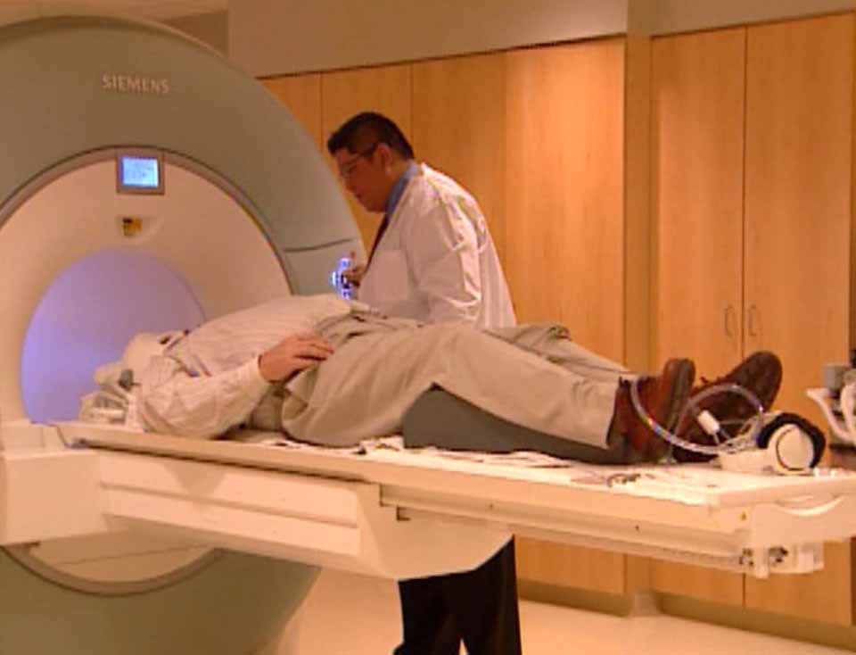
Figure 1: Patient Brent Boyd entering an MRI scanner. High-res (TIF) version (Right-click and Save As) |
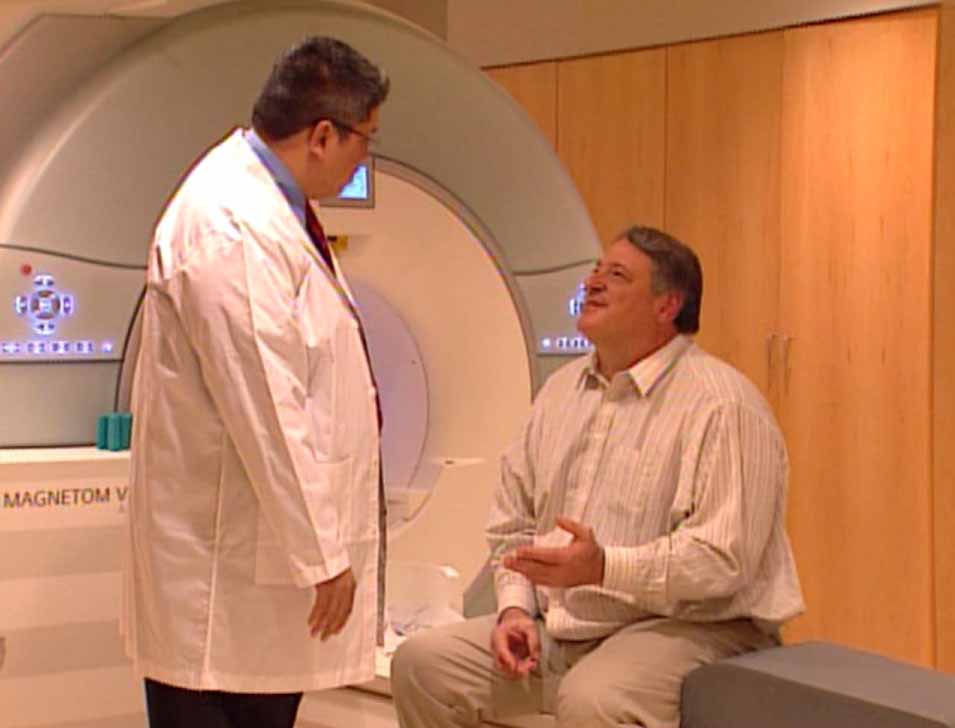
Figure 2: Dr. Alexander Lin talking with patient Brent Boyd. High-res (TIF) version (Right-click and Save As) |
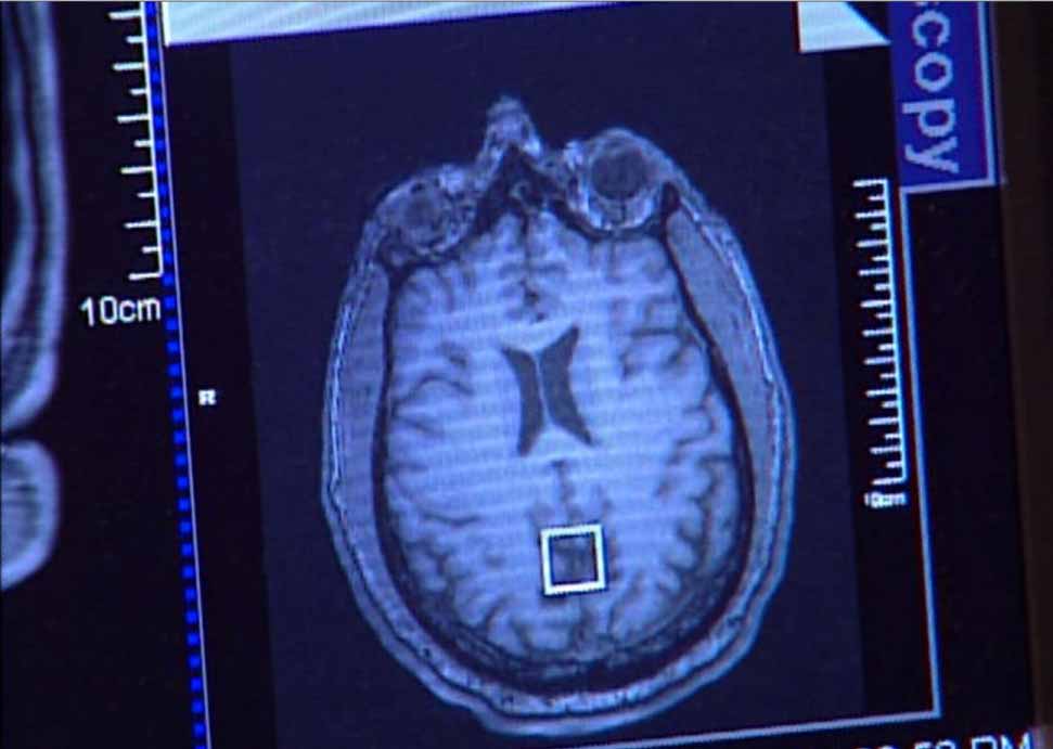
Figure 3: A head MR image. High-res (TIF) version (Right-click and Save As) |
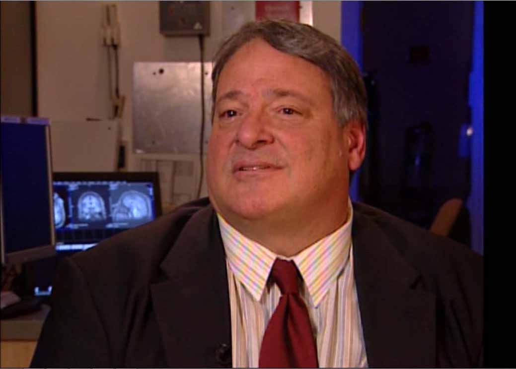
Figure 4: Former NFL player Brent Boyd. High-res (TIF) version (Right-click and Save As) |
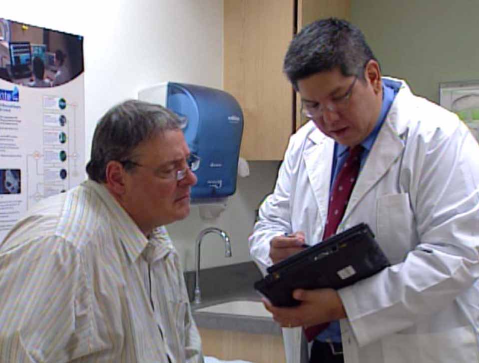
Figure 5: Dr. Alexander Lin and Brent Boyd talking in a consult room. High-res (TIF) version (Right-click and Save As) |
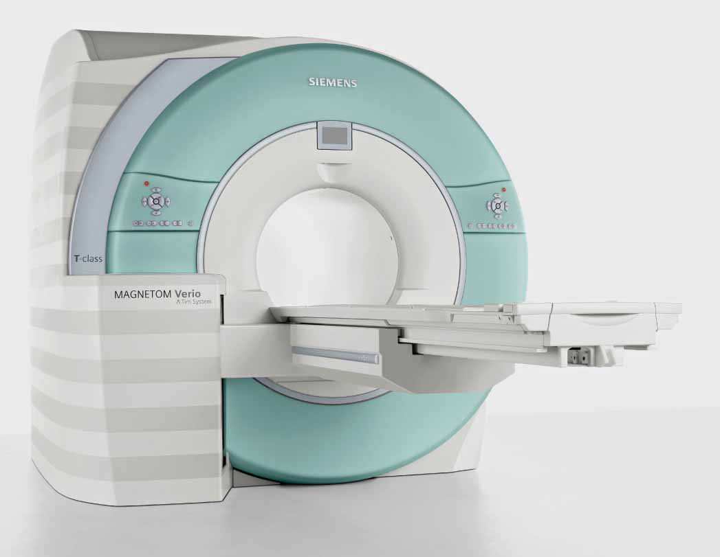
Verio 1: A photograph of the Siemens TIM Verio MR scanner. High-res (TIF) version (Right-click and Save As) |
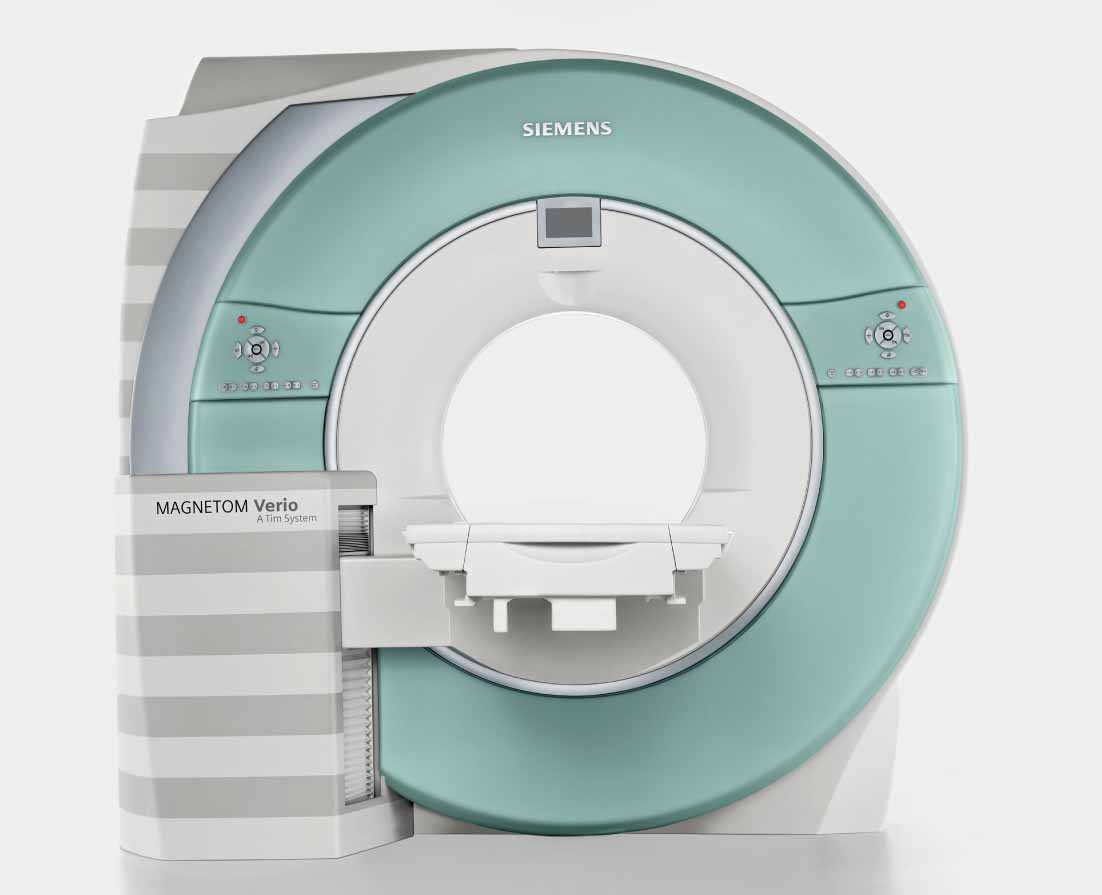
Verio 2: A photograph of the Siemens TIM Verio MR scanner. High-res (TIF) version (Right-click and Save As) |
 PDF
PDF