RSNA Press Release
- N-acetylaspartate (NAA) levels in the part of the brain associated with pleasure and pain are reduced in smokers in direct relationship to how much they've smoked.
- Decreased NAA levels in the brain have been associated with mood disorders and substance abuse.
- A correlation was found between increased total creatine levels in the frontal lobes of the brain and likelihood of smoking relapse.
- NAA levels normalized after six months of smoking cessation.
Smoking Changes Brain Chemistry
Released: November 28, 2006
| Media Contacts: | ||
| RSNA Media Relations: | (630) 590-7762 | |
| Maureen Morley (630) 590-7754 mmorley@rsna.org |
||
CHICAGO — Chronic smoking affects nerve cells and alters the chemical makeup of the brain, according to research presented today at the annual meeting of the Radiological Society of North America (RSNA).
"This is the first imaging study to focus on the relationship between brain metabolites and nicotine dependence," said Okan Gür, M.D., from the Department of Radiology at the University of Bonn in Germany.
Dr. Gür and colleagues used proton magnetic resonance spectroscopy (MRS) to study 21 men and 22 women, age 21 to 59, in a smoking cessation program two weeks after quitting and again six months later. Patients were encouraged to use nicotine patches during the initial six weeks of smoking cessation; however, only 36 of the patients complied.
Proton MRS is able to measure brain metabolism at the cellular level and can provide detailed chemical data about the brain's metabolites, which are involved in many physical and chemical processes within the body.
The researchers compared the data collected from the smokers to proton MRS data collected from 35 age- and gender-matched healthy controls. The results showed that the nicotine-dependent patients had significantly decreased concentrations of the amino acid N-acetylaspartate (NAA) in the anterior cingulate cortex (ACC), the part of the brain that processes pleasure and pain. The decreased NAA levels were evident regardless of whether or not the patient used a nicotine patch and correlated directly with the patient's smoking history: the greater the number of pack years (one pack per day for one year equals one pack year), the lower the NAA level.
"The ACC is involved in mediating conditioned reinforcement, craving and relapsing behavior in addiction," said study co-author Christian G. Schütz, M.D., M.P.H., from the Department of Psychiatry at the University of Bonn. "Lower NAA levels have been implicated as indicators of neuronal or axonal dysfunction."
Reduced NAA levels have been reported for a number of psychiatric and mood disorders, including schizophrenia, dementia and bipolar disorder, as well as in cases of substance abuse, particularly alcohol dependence.
Choline concentrations in the ACC were slightly lower in the smokers compared with the nonsmokers, and lower still in the female smokers compared with the male smokers. Choline is significantly involved in cell membrane metabolism, which is essential for cardiac and brain function. Reduced choline levels can be a precursor to the breakdown of cell membranes.
Concentration of total creatine (tCr) levels in the frontal lobes was typically higher in the smokers who did not use patches compared to those who did. The metabolite tCr plays an important role in supplying energy to the muscle cells. Higher tCr levels have been associated with stimulant use. Furthermore, the researchers found that high tCr levels predicted a higher likelihood of relapse.
Upon follow-up after six months, the researchers found that most metabolite concentrations, including that of NAA, had normalized in the 25 ex-smokers who did not relapse.
"These findings further emphasize the importance of quitting smoking," Dr. Gür said. "The degree of reduction of NAA in the ACC depends on the amount of tobacco consumed over time, but it appears to normalize after smoking cessation."
Co-authors are Hans H. Schild, M.D., Wolfgang Block, Ph.D., Frank Träber, Ph.D., and Wolfgang Maier, M.D.
# # #
RSNA is an association of more than 40,000 radiologists, radiation oncologists, medical physicists and related scientists committed to promoting excellence in radiology through education and by fostering research, with the ultimate goal of improving patient care. The Society is based in Oak Brook, Ill.
Editor's note: The data in these releases may differ from those in the printed abstract and those actually presented at the meeting, as researchers continue to update their data right up until the meeting. To ensure you are using the most up-to-date information, please call the RSNA Newsroom at (312) 949-3233.
| Abstract: |
Images (.JPG format)
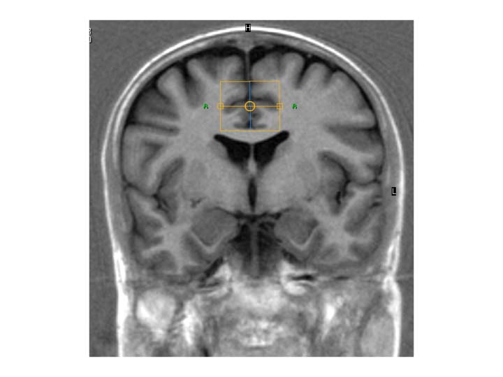 Figure 1a. High-res (TIF) version (Right-click and Save As) |
 Figure 1b. High-res (TIF) version (Right-click and Save As) |
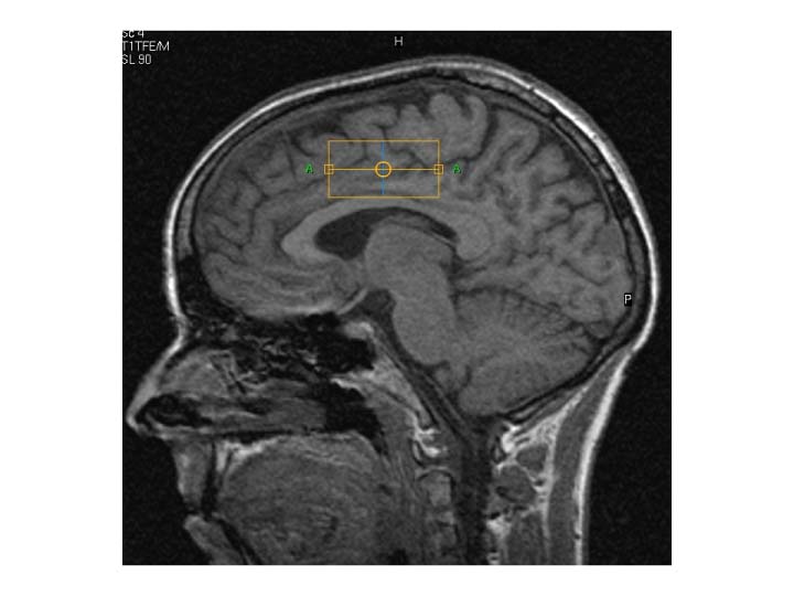 Figure 1c. High-res (TIF) version (Right-click and Save As) |
|
| First Volumes of interest (VOI) of 45 x 28 x 22 mm were placed superior to the corpus callosum, covering gyrus cinguli anterior in both hemispheres (see yellow boxes in figures). | |||
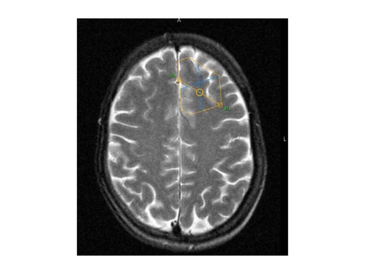 Figure 2a. High-res (TIF) version (Right-click and Save As) |
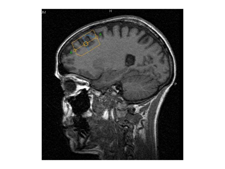 Figure 2b. High-res (TIF) version (Right-click and Save As) |
||
| Second VOI of 40 x 30 x 25 mm was placed anterior to the sulcus precentralis in the left dorsolateral frontal cortex, subjacent white matter, and the anterior cingulate cortex (see yellow boxes in figures). | |||
 Figure 3. Normal distribution of metabolites in cerebral H-MR spectra High-res (TIF) version (Right-click and Save As) |
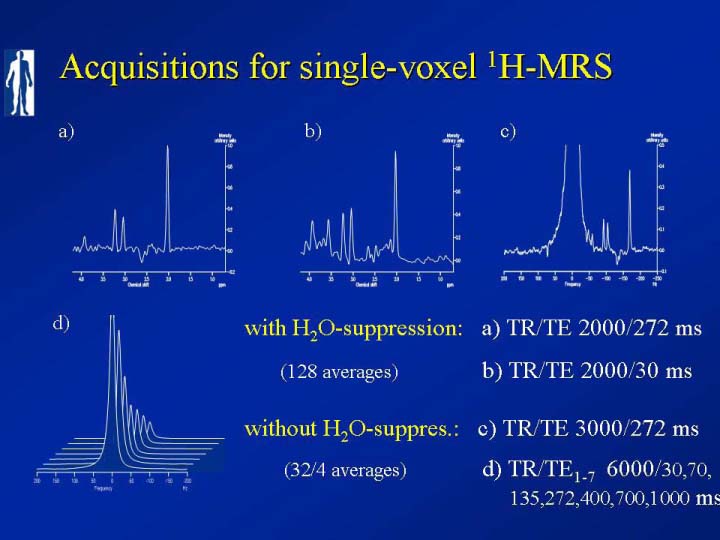 Figure 4. Acquisitions for single voxel H-MRS High-res (TIF) version (Right-click and Save As) |
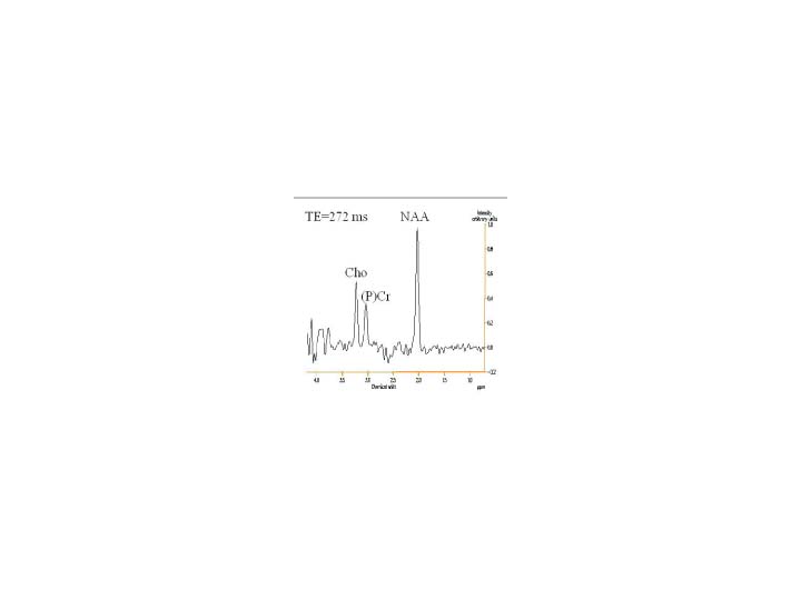 Figure 5. Graph - Single-voxel H-MRS of the cingulum TE272 High-res (TIF) version (Right-click and Save As) |
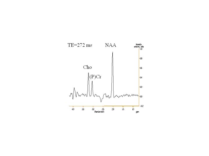 Figure 6. Graph - Single-voxel H-MRS of the frontal cortex TE272 High-res (TIF) version (Right-click and Save As) |
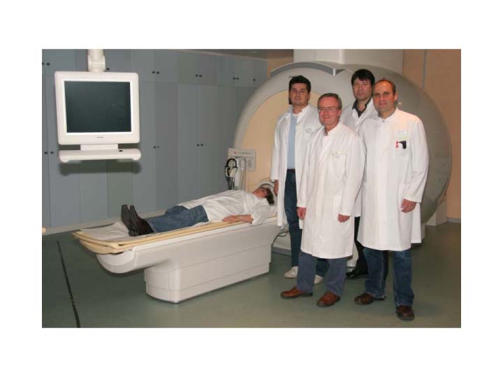 Figure 7. The research team and equipment High-res (TIF) version (Right-click and Save As) |
|||
 PDF
PDF