Stronger Thigh Muscles May Prevent Knee Replacement Surgery
Released: November 27, 2023
At A Glance
- Stronger quadriceps muscles, relative to the hamstrings, may lower the risk of total knee replacement.
- Researchers evaluated thigh muscle volume in 134 participants and found a higher ratio of quadricep-hamstring volume was significantly associated with lower odds of total knee replacement.
- The results suggest that training programs that strengthen the quadriceps compared to the hamstrings may be beneficial.
- RSNA Media Relations
1-630-590-7762
media@rsna.org - Linda Brooks
1-630-590-7738
lbrooks@rsna.org - Imani Harris
1-630-481-1009
iharris@rsna.org
CHICAGO — Stronger quadriceps muscles, relative to the hamstrings, may lower the risk of total knee replacement, according to research being presented today at the annual meeting of the Radiological Society of North America (RSNA). Researchers said the findings could inform strength-training programs for people with advanced arthritis in the knee.

Advanced knee osteoarthritis is a major cause of pain and disability worldwide. In the U.S. alone, 14 million adults have symptomatic knee osteoarthritis, and more than half of those diagnosed are projected to eventually undergo total knee replacement surgery.
While stronger muscle groups are generally understood to be associated with a lower rate of total knee replacement, their relative importance is not well established. Of particular interest is the relationship between the extensors and the hamstrings, the two most important muscle groups in the knee.
The extensors, the muscles on the front of the thigh commonly referred to as the quadriceps, are the strongest muscle group in the body and have essential influence on gait, other activities and biomechanics. The muscles around the back of the thigh known as the hamstrings are responsible for extension of the hip and flexion of the knee, making them equally essential for physical activity.
“The two muscle groups act as counter forces, and the balance between them enables a wide range of activities while protecting the knee joint,” said study lead author Upasana Upadhyay Bharadwaj, M.D., from the University of California, San Francisco (UCSF). “An imbalance, in addition to other factors, leads to a change in the biomechanics resulting in the progression of osteoarthritis.”
Dr. Upadhyay Bharadwaj and colleagues evaluated thigh muscle volume in 134 participants from the Osteoarthritis Initiative, a nationwide study sponsored by the National Institutes of Health. They compared 67 patients who underwent total knee replacement of a single knee with 67 control participants who had not undergone knee replacement. The cases and controls were matched for variables including age and gender.
The researchers obtained 3T MRI of the thigh at the time of surgery. They also evaluated MRI findings from two years and four years before the surgery. They used a previously trained deep-learning model to segment and compute volumes of the muscles of the thigh—measures that are tedious to compute manually.
Comparing patients who had total knee replacement with the control group, a higher ratio of quadriceps to hamstring volume was significantly associated with lower odds of total knee replacement. Higher volumes of hamstrings and gracilis, a long, thin muscle on the inside of the thigh, were also linked with lower odds of total knee replacement.
“Our study shows that in addition to strong muscles individually, larger extensor muscle groups—relative to hamstring muscle groups—are significantly associated with lower odds of total knee replacement surgery in two to four years,” Dr. Upadhyay Bharadwaj said.
The study findings have implications for both the interpretation of imaging exams and clinical management. The results suggest that training programs that strengthen the quadriceps in relation to the hamstrings may be beneficial.
“Although we presume that overall muscle volume is important as a surrogate marker for muscle strength, the ratio, hence the balance, between extensor and hamstring muscles may be more important and significantly associated with lower odds of total knee replacement,” Dr. Upadhyay Bharadwaj said.
Although the study focused on people with arthritis, the findings may also help inform strength training for a wider segment of the population.
“While these results are essential for targeted therapy in a population at risk for osteoarthritis, even the general public can benefit from our results to preventively incorporate appropriate strengthening exercises,” Dr. Upadhyay Bharadwaj said.
Co-authors are John A. Lynch, Ph.D., Gabby B. Joseph, Ph.D., and Thomas M. Link, M.D., Ph.D.
Note: Copies of RSNA 2023 news releases and electronic images will be available online at RSNA.org/press23.
RSNA is an association of radiologists, radiation oncologists, medical physicists and related scientists promoting excellence in patient care and health care delivery through education, research and technologic innovation. The Society is based in Oak Brook, Illinois. (RSNA.org)
Editor’s note: The data in these releases may differ from those in the published abstract and those actually presented at the meeting, as researchers continue to update their data right up until the meeting. To ensure you are using the most up-to-date information, please call the RSNA Newsroom at 1-312-791-6610.
For patient-friendly information on musculoskeletal imaging, visit RadiologyInfo.org.
Video (MP4):

Video. Upasana Upadhyay Bharadwaj, M.D., discusses her research on how stronger quadriceps muscles relative to the hamstrings, may lower the risk of total knee replacement.
Download MP4
(Right-click and Save As)
Images (JPG, TIF):
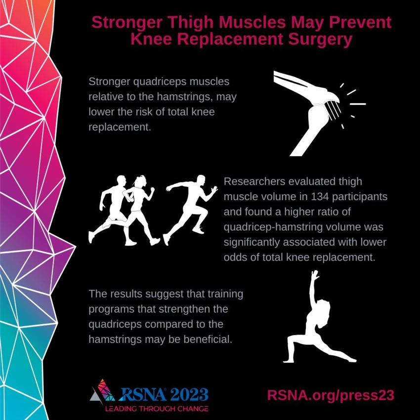
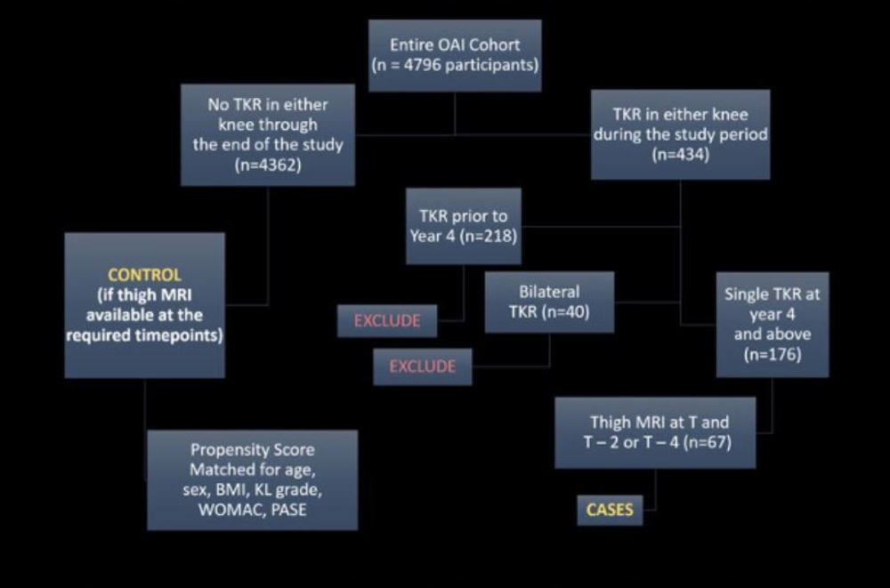
Figure 1. Participant selection of cases and matched controls from the Osteoarthritis Initiative.
High-res (TIF) version
(Right-click and Save As)
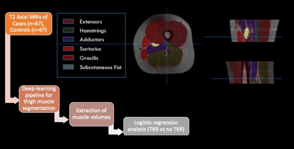
Figure 2. Study design for analysis of MRI with visualization of the deep-learning-based 3D segmentation model.
High-res (TIF) version
(Right-click and Save As)
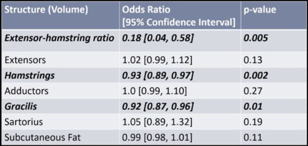
Figure 3. Results of logistic regression analysis with odds ratio (total knee replacement vs no total knee replacement) and confidence intervals reported for each fixed effect.
High-res (TIF) version
(Right-click and Save As)
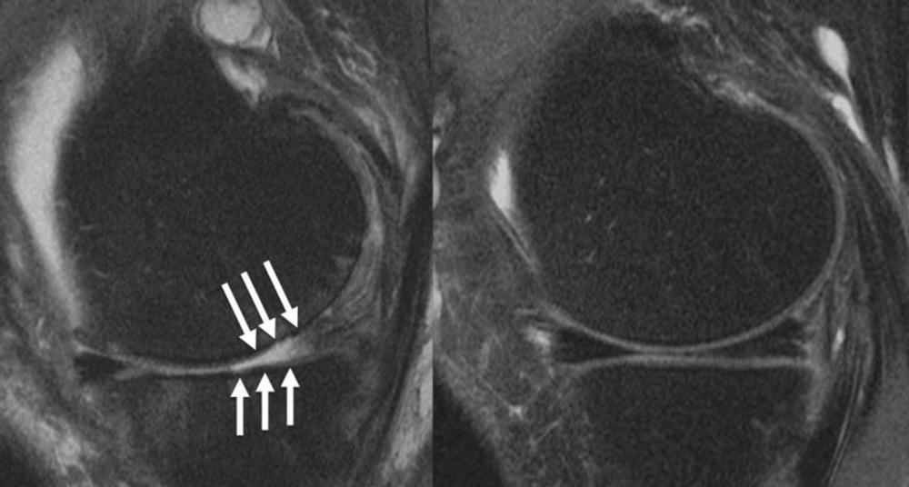
Figure 4. : Knee joint of a patient showing (A) severe cartilage defects and (B) intact knee joint.
High-res (TIF) version
(Right-click and Save As)
Additional Resources