New Studies Show Brain Impact of Youth Football
Released: November 27, 2017
At A Glance
- Two new studies revealed changes in the default mode network in the brains of young football players.
- One study used machine learning to predict which players had high- and low-impact exposure based on fMRI results.
- A second study showed DMN changes may be dependent on a player's previous concussion history.
- RSNA Media Relations
1-630-590-7762
media@rsna.org - Maureen Morley
1-630-590-7754
mmorley@rsna.org - Linda Brooks
1-630-590-7738
lbrooks@rsna.org

CHICAGO — School-age football players with a history of concussion and high impact exposure undergo brain changes after one season of play, according to two new studies conducted at UT Southwestern Medical Center in Dallas and Wake Forest Baptist Medical Center in Winston-Salem and presented today at the annual meeting of the Radiological Society of North America (RSNA).
Both studies analyzed the default mode network (DMN), a network of brain regions that is active during wakeful rest. Changes in the DMN are observed in patients with mental disorders. Decreased connectivity within the network is also associated with traumatic brain injury.
"The DMN exists in the deep gray matter areas of the brain," explained Elizabeth M. Davenport, Ph.D., a postdoctoral researcher in the Advanced NeuroScience Imaging Research (ANSIR) lab at UT Southwestern's O'Donnell Brain Institute. "It includes structures that activate when we are awake and engaging in introspection or processing emotions, which are activities that are important for brain health."
In the first study, researchers studied youth football players without history of concussion to identify the effect of repeated subconcussive impacts on the DMN.

"Over a season of football, players are exposed to numerous head impacts. The vast majority of these do not result in concussion," said Gowtham Krishnan Murugesan, a Ph.D. student in biomedical engineering and member of the ANSIR lab. "This work adds to a growing body of literature indicating that subconcussive head impacts can have an effect on the brain. This is a highly understudied area at the youth and high school level."
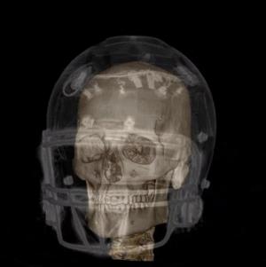
For the study, 26 youth football players (ages 9-13) were outfitted with the Head Impact Telemetry System (HITS) for an entire football season. HITS helmets are lined with accelerometers or sensors that measure the magnitude, location and direction of impacts to the head. Impact data from the helmets were used to calculate a risk of concussion exposure for each player.
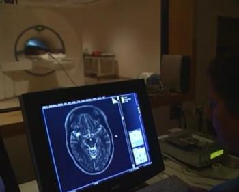
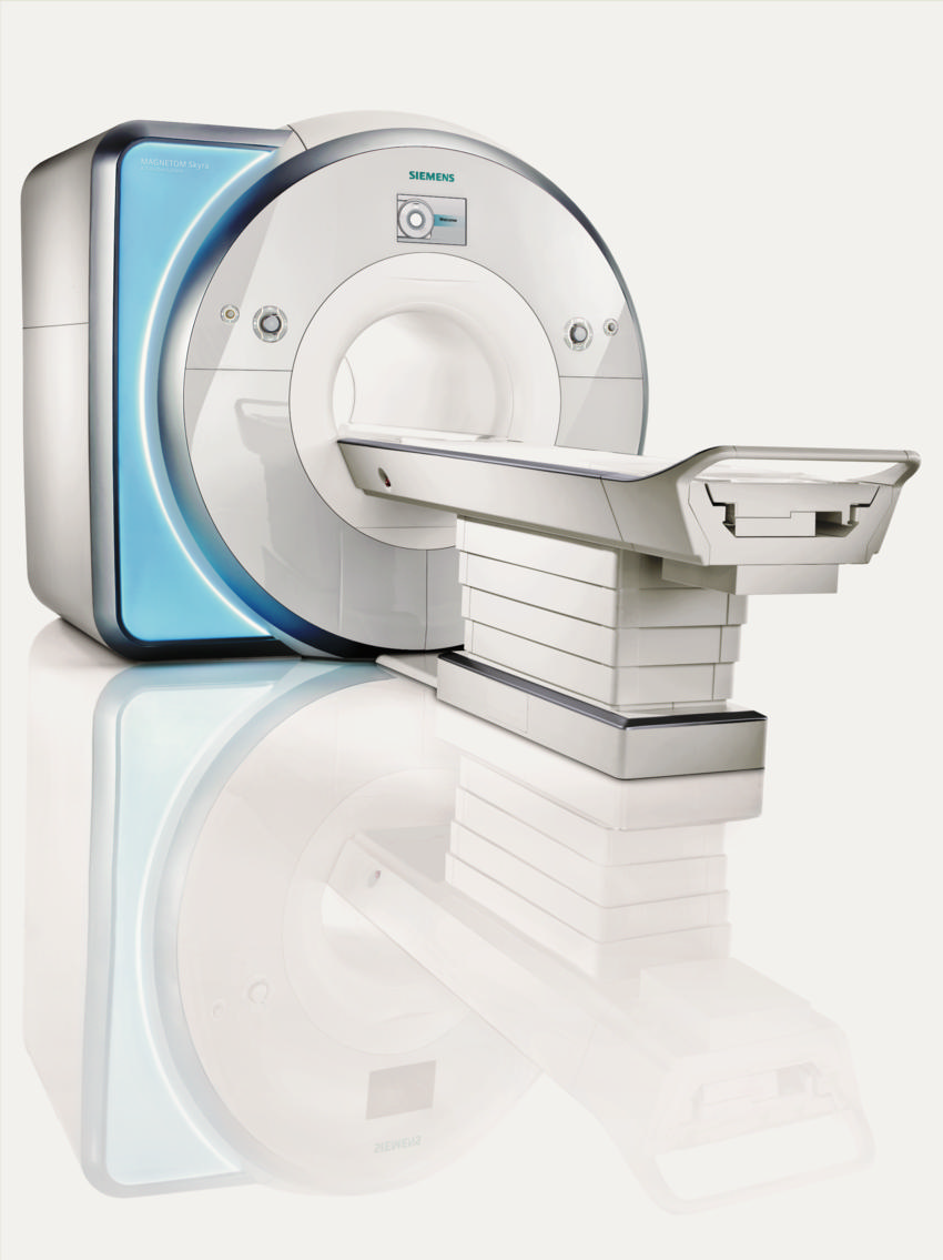
Players were equally divided into high and low concussion exposure groups. Players with a history of concussion were excluded. A third group of 13 non-contact sport controls was established. Pre- and post-season resting functional MRI (fMRI) scans were performed on all players and controls, and connectivity within the DMN sub-components was analyzed.
The researchers used machine learning to analyze the fMRI data. Machine learning is a type of artificial intelligence that allows computers to perform analyses based on existing relationships of data.
"Machine learning has a lot to add to our research because it gives us a fresh perspective and an ability to analyze the complex relationships within the data," said Murugesan. "Our results suggest an increasing functional change in the brain with increasing head impact exposure."
Five machine learning classification algorithms were used to predict whether players were in the high-exposure, low-exposure or non-contact groups based on the fMRI results. The algorithm discriminated between high-impact exposure and non-contact with 82 percent accuracy, and low-impact exposure and non-contact with 70 percent accuracy. The results suggest an increasing functional change with increasing head-impact exposure.
"The brains of these youth and adolescent athletes are undergoing rapid maturation in this age range. This study demonstrates that playing a season of contact sports at the youth level can produce neuroimaging brain changes, particularly for the DMN," Murugesan said.
In the second study, 20 high school football players (median age 16.9) wore helmets outfitted with HITS for a season. Of the 20 players, five had experienced at least one concussion, and 15 had no history of concussion.
Before and following the season, the players underwent an eight-minute magnetoencephalography (MEG) scan, which records and analyzes the magnetic fields produced by brain activity. Researchers then analyzed the MEG power associated with the eight brain regions of the DMN.
Post-season, the five players with a history of concussion had significantly lower connectivity between DMN regions. Players with no history of concussion had, on average, an increase in DMN connectivity.
The results demonstrate that concussions from previous years can influence the changes occurring in the brain during the current season, suggesting that there are longitudinal effects of concussion that affect brain function.
"The brain's default mode network changes differently as a result of previous concussion," Dr. Davenport said. "Previous concussion seems to prime the brain for additional changes. Concussion history may be affecting the brain's ability to compensate for subconcussive impacts."
Both researchers said larger data sets, longitudinal studies that follow young football players and research that combines both MEG and fMRI are needed to better understand the complex factors involved in concussions.
Murugesan co-authors are Afarin Famili, Elizabeth M. Davenport, Ph.D., Ben Wagner, B.M., Christopher T. Whitlow, M.D., Ph.D., Joel Stitzel, Jillian Urban, Joseph A. Maldjian, M.D., and Albert Montillo, Ph.D.
Davenport co-authors are Jillian Urban, Ben Wagner, B.M., Mark A. Espeland, Ph.D., Alexander K. Powers, M.D., Christopher T. Whitlow, M.D., Ph.D., Joel Stitzel, and Joseph A. Maldjian, M.D. This work is part of several NIH-funded studies of youth and high school football, with additional funding from the Childress Institute for Pediatric Trauma. Data collection performed by researchers at Wake Forest University, and data analysis done by researchers at UT Southwestern.
Note: Copies of RSNA 2017 news releases and electronic images will be available online at RSNA.org/press17 beginning Monday, Nov. 27.
RSNA is an association of over 54,000 radiologists, radiation oncologists, medical physicists and related scientists, promoting excellence in patient care and health care delivery through education, research and technologic innovation. The Society is based in Oak Brook, Ill. (RSNA.org)
Editor’s note: The data in these releases may differ from those in the published abstract and those actually presented at the meeting, as researchers continue to update their data right up until the meeting. To ensure you are using the most up-to-date information, please call the RSNA Newsroom at 1-312-791-6610.
For patient-friendly information on brain imaging, visit RadiologyInfo.org.
Video clips (.mp4 format)
Video 1. Animation showing how the HIT system accelerometers fit around the player's head. The springs ensure the sensors remain in contact with the head, measuring head acceleration not helmet acceleration.
Download.mp4
(Right-click and Save As)
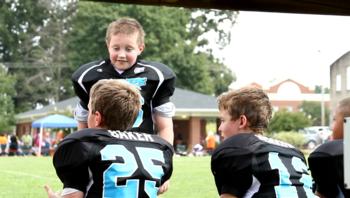
Video 2. B-roll footage of adolescents playing football using helmets equipped with HITS.
Download.mp4
(Right-click and Save As)
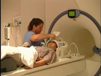
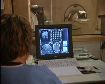
Video 3. Radiologic technologist positions patient for fMRI exam
Download.mp4
(Right-click and Save As)

Video 7. Dr. Elizabeth M. Davenport and Gowtham Krishnan Murugesan discuss their research on brain impact in youth football.
Download.mp4
(Right-click and Save As)

Video 8. Dr. Elizabeth M. Davenport and Gowtham Krishnan Murugesan discuss default mode network and why it is important.
Download.mp4
(Right-click and Save As)

Video 9. Dr. Elizabeth M. Davenport and Gowtham Krishnan Murugesan discuss the takeaway message of their research.
Download.mp4
(Right-click and Save As)

Video 10. Dr. Elizabeth M. Davenport and Gowtham Krishnan Murugesan discuss surprising findings from their research.
Download.mp4
(Right-click and Save As)

Video 11. Dr. Elizabeth M. Davenport and Gowtham Krishnan Murugesan give their advice to parents of children and adolescents who want to play football.
Download.mp4
(Right-click and Save As)
Images (.JPG and .TIF format)
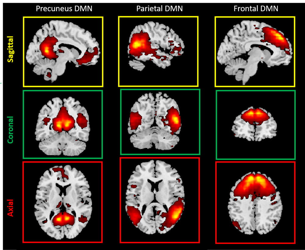
Figure 1. Examples of Default Mode Network (DMN) subcomponents examined in this study
High-res (TIF) version
(Right-click and Save As)

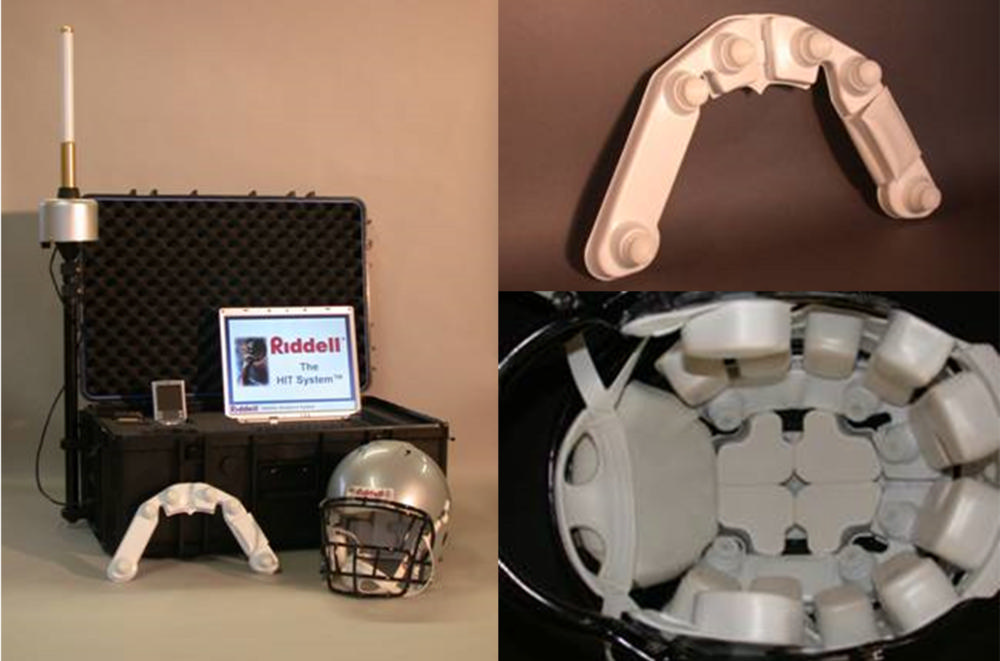
Figure 3. Riddell HIT System with a sensor array and helmet
High-res (TIF) version
(Right-click and Save As)

Figure 4. Vectors representing head impacts recorded with the HIT System
High-res (TIF) version
(Right-click and Save As)
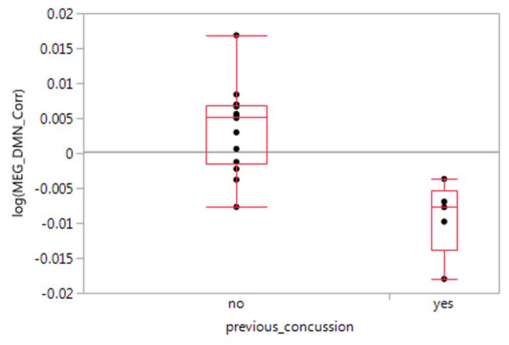
Figure 5. MEG DMN correlation between players with previous concussion and no concussion
High-res (TIF) version
(Right-click and Save As)
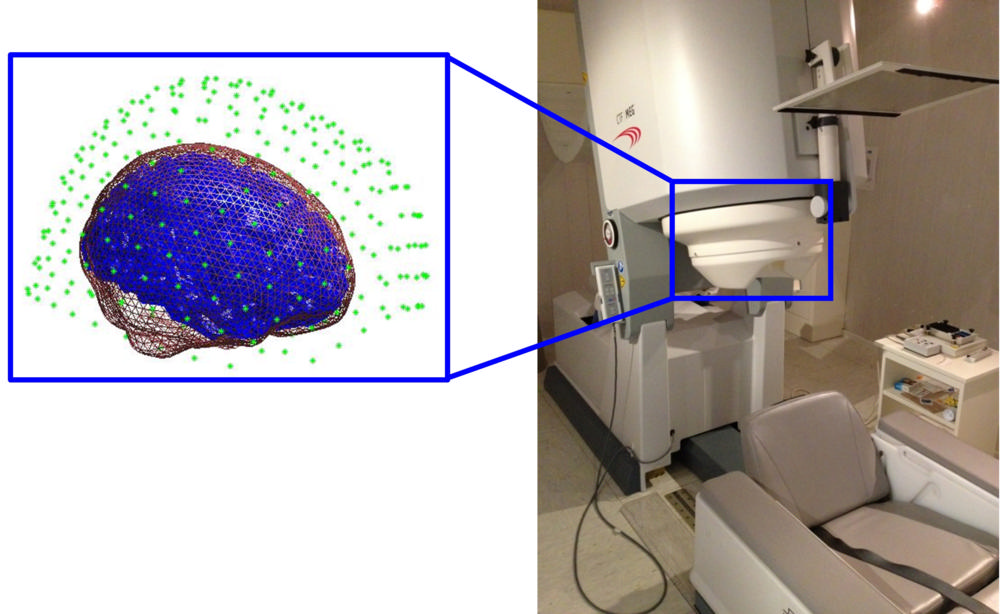
Figure 6. Magnetoencephalography (MEG) equipment. The green dots represent the fit of the sensors around a patient's brain.
High-res (TIF) version
(Right-click and Save As)