Study Finds Association Between Coronary Artery Plaque and Liver Disease
Released: November 04, 2014
At A Glance
- Researchers looked at associations between high-risk coronary artery plaque and non-alcoholic fatty liver disease (NAFLD).
- High-risk coronary artery plaque was seen in 59.3 percent of patients with NAFLD, compared to only 19 percent of those without NAFLD.
- NAFLD is the most common liver disease, with an estimated prevalence of 20 percent to 30 percent in the general population.
- RSNA Media Relations
1-630-590-7762
media@rsna.org - Linda Brooks
1-630-590-7738
lbrooks@rsna.org - Emma Day
1-630-590-7791
eday@rsna.org
OAK BROOK, Ill. — Researchers using coronary computed tomography angiography (CCTA) have found a close association between high-risk coronary artery plaque and a common liver disease. The study, published online in the journal Radiology, found that a single CT exam can detect both conditions.
Previous research has shown that CCTA can detect high-risk coronary artery plaque, or plaque prone to life-threatening ruptures. For the new study, researchers looked at associations between high-risk plaque and non-alcoholic fatty liver disease (NAFLD), a condition characterized by abnormal liver function that is not associated with excessive alcohol consumption. NAFLD is the most common liver disease, with an estimated prevalence of 20 percent to 30 percent in the general population.
"As it is known that atherosclerosis is linked to inflammation, our next step was to look for an association of high-risk plaques with other systemic inflammatory conditions such as NAFLD," said the study's lead author, Stefan B. Puchner, M.D., from Massachusetts General Hospital and Harvard Medical School in Boston and the Medical University of Vienna, Austria. "Interestingly, both pathologies can be detected in a single CT examination."
The researchers drew patients from a large trial focusing on the use of CCTA in people who had come to the emergency department with acute chest pain. The patients underwent both non-enhanced CT to assess coronary calcium, a marker of atherosclerosis, and contrast-enhanced CCTA. Readers assessed the CCTA images for signs of high-risk plaque.
Overall, 182 of the 445 patients in the study, or 40.9 percent, had CT evidence of NAFLD. High-risk plaque was seen in 59.3 percent of patients with NAFLD, compared to only 19 percent of those without NAFLD. The association between NAFLD and high-risk plaque persisted after adjusting for the extent and severity of coronary atherosclerosis and traditional risk factors.
The results suggest that high-risk plaque and NAFLD are both part of the same systemic disease process, the metabolic syndrome.
"The clinical implications could include a wider assessment of NAFLD using the non-contrast cardiac CT scans that are commonly performed prior to the CTA," he said. "Further, the additional assessment of NAFLD with CT could improve the risk stratification of patients with suspected coronary artery disease, as our results show that the presence of NAFLD is associated with high-risk coronary plaque independent of traditional risk factors and severity of coronary artery disease."
The researchers plan to extend the study outside of the emergency department setting to see if the results apply to other categories of the general population. They also hope to learn more about why NAFLD is so prevalent among people with advanced high-risk coronary atherosclerosis. One prevailing theory is that both conditions are a consequence of systemic inflammation, an inflammatory state that affects multiple organs and can lead to life-threatening conditions.
"The aim will be to further investigate and understand, with the help of CCTA, the interplay between advanced atherosclerosis and NAFLD as part of a complex systemic inflammatory condition," Dr. Puchner said.
"High-Risk Coronary Plaque at Coronary CT Angiography Is Associated with Nonalcoholic Fatty Liver Disease, Independent of Coronary Plaque and Stenosis Burden." Collaborating with Dr. Puchner were Michael T. Lu, M.D., Thomas Mayrhofer, Ph.D., Ting Liu, M.D., Amit Pursnani, M.D., Brian B. Ghoshhajra, M.D., M.B.A., Quynh A. Truong, M.D., M.P.H., Stephen D. Wiviott, M.D., Jerome L. Fleg, M.D., Udo Hoffmann, M.D., M.P.H., and Maros Ferencik, M.D., Ph.D.
Radiology is edited by Herbert Y. Kressel, M.D., Harvard Medical School, Boston, Mass., and owned and published by the Radiological Society of North America, Inc. (http://radiology.rsna.org/)
RSNA is an association of more than 54,000 radiologists, radiation oncologists, medical physicists and related scientists, promoting excellence in patient care and health care delivery through education, research and technologic innovation. The Society is based in Oak Brook, Ill. (RSNA.org)
For patient-friendly information on CCTA, visit RadiologyInfo.org.
Press Resources:
Images (.JPG and .TIF format)
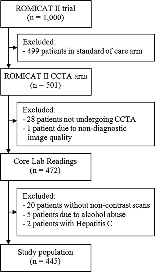
Figure 1. Flowchart demonstrates study popula¬tion enrollment, both exclusion and inclusion. CCTA = coronary CT angiography.
High-res (TIF) version
(Right-click and Save As)
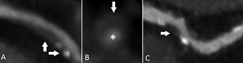
Figure 2. Coronary CT angiography images demonstrate examples of high-risk plaque features. A, Image was obtained in a 63-year-old man with partially calcified plaque, positive remodeling (vertical arrow), and spotty calcium (horizontal arrow). B, A cross-sectional view of a noncalcified plaque in a 65-year-old man demonstrates a napkin-ring sign with a central low-attenuation area, surrounded by a peripheral rim of higher attenuation (arrow) next to the lumen (∗). C, Image in a 60-year-old woman with partially calcified plaque demonstrates a low CT number in the midportion (arrow).
High-res (TIF) version
(Right-click and Save As)
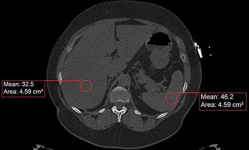
Figure 3. Axial nonenhanced CT image in a 62-year-old man demonstrates attenuation measurements of the liver and spleen. Image shows a diffuse fat accumulation in the liver, with a mean liver attenuation of 32.5 HU and a mean spleen attenuation of 46.2 HU.
High-res (TIF) version
(Right-click and Save As)