Elevated MRI Enhancement Ups Cancer Risk in Women with Very Dense Breasts
Released: August 08, 2023
At A Glance
- Researchers trained a machine learning model using dynamic contrast-enhanced MRI exams from 4,553 participants to assess breast cancer risk in women with extremely dense breasts.
- Cancer occurrence was greater in women with higher volumes of enhancing parenchyma on breast MRI.
- The findings suggest potential to tailor supplemental MRI screening to individual women by anticipating interval cancers.
- RSNA Media Relations
1-630-590-7762
media@rsna.org - Linda Brooks
1-630-590-7738
lbrooks@rsna.org - Imani Harris
1-630-481-1009
iharris@rsna.org
OAK BROOK, Ill. — A machine learning model found that background parenchymal enhancement (BPE) on breast MRI is an indicator of breast cancer risk in women with extremely dense breasts, according to a study published in Radiology, a journal of the Radiological Society of North America (RSNA).
Women with extremely dense breasts are at a three- to six-times higher risk of developing breast cancer compared to women who have fatty breasts. Since mammography is less sensitive in detecting early-stage breast cancer in women with dense breasts, women between the ages of 50 and 75 years with dense breasts may benefit from additional MRI screening.
Another breast cancer risk factor is BPE, which is the level of normal fibroglandular tissue that enhances on breast MRI. However, not much is known about how BPE compares to other more established clinical risk factors of breast cancer, such as age, body mass index (BMI) family history, and breast density.
"Thus far, studies on breast cancer risk factors have typically focused on women at high lifetime risk of developing breast cancer," said study co-author Kenneth G. A. Gilhuijs, Ph.D., from the Department of Radiology at the University Medical Center Utrecht in the Netherlands. "This is the first study that we know of that demonstrates an association between background parenchymal enhancement and occurrence of breast cancer in women with extremely dense breasts."

To determine how much BPE is an indicator of breast cancer risk, the researchers used dynamic contrast-enhanced MRI exams from 4,553 participants in the Dense Tissue and Early Breast Neoplasm Screening (DENSE) Trial, a large multi-institutional study based in the Netherlands, to develop a deep learning model to automatically identify the fibroglandular tissue. The MRI exams were performed every two years in eight hospitals in the Netherlands between December 2011 and January 2016.
After adjusting for age, BMI and BPE, the researchers found that breast cancer occurrence was greater in women with higher volumes of enhancing parenchyma compared to women with low volumes of enhancing parenchyma.
Of the 4,553 women included in the study, 122 were diagnosed with breast cancer. Roughly 63% of them were diagnosed after the first round of screening. An average cancer detection time of 24 months was associated with the remaining women diagnosed with breast cancer.
"Parenchyma does not enhance uniformly on MRI," Dr. Gilhuijs said. "This method calculates all the different subvolumes at which the parenchyma enhances and sorts them from high to low."
The researchers point out that while the implementation of supplemental MRI screening in women with dense breasts will result in fewer interval cancers—which are breast cancers that are diagnosed in between routine mammography screenings—it will also further strain radiologist workloads. Developing more personalized strategies to deal with the added number of screenings may help alleviate the strain on the healthcare field.
"Our study is a first step in a direction to further tailor the frequency of supplemental MRI screening to individual women with dense breasts, focusing not only on breast density as a main risk factor but also on other properties of the breast established from a first screening MRI," Dr. Gilhuijs said.
"Assessing Quantitative Parenchymal Features at Baseline Dynamic Contrast-enhanced MRI and Cancer Occurrence in Women with Extremely Dense Breasts." Collaborating with Dr. Gilhuijs were Hui Wang, M.D., Bas H. M. van der Velden, Ph.D., Erik Verburg, Ph.D., Marije F. Bakker, Ph.D., Ruud M. Pijnappel, M.D., Ph.D., Wouter B. Veldhuis, M.D., Ph.D., and Carla H. van Gils, Ph.D.
In 2023, Radiology is celebrating its 100th anniversary with 12 centennial issues, highlighting Radiology's legacy of publishing exceptional and practical science to improve patient care.
Radiology is edited by Linda Moy, M.D., New York University, New York, N.Y., and owned and published by the Radiological Society of North America, Inc. (https://pubs.rsna.org/journal/radiology)
RSNA is an association of radiologists, radiation oncologists, medical physicists and related scientists promoting excellence in patient care and health care delivery through education, research and technologic innovation. The Society is based in Oak Brook, Illinois. (RSNA.org)
For patient-friendly information on breast imaging, visit RadiologyInfo.org.
Images (JPG, TIF):
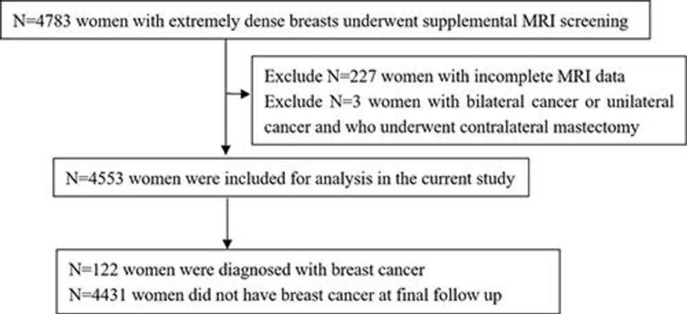
Figure 1. Flowchart of selection of women included in this study.
High-res (TIF) version
(Right-click and Save As)
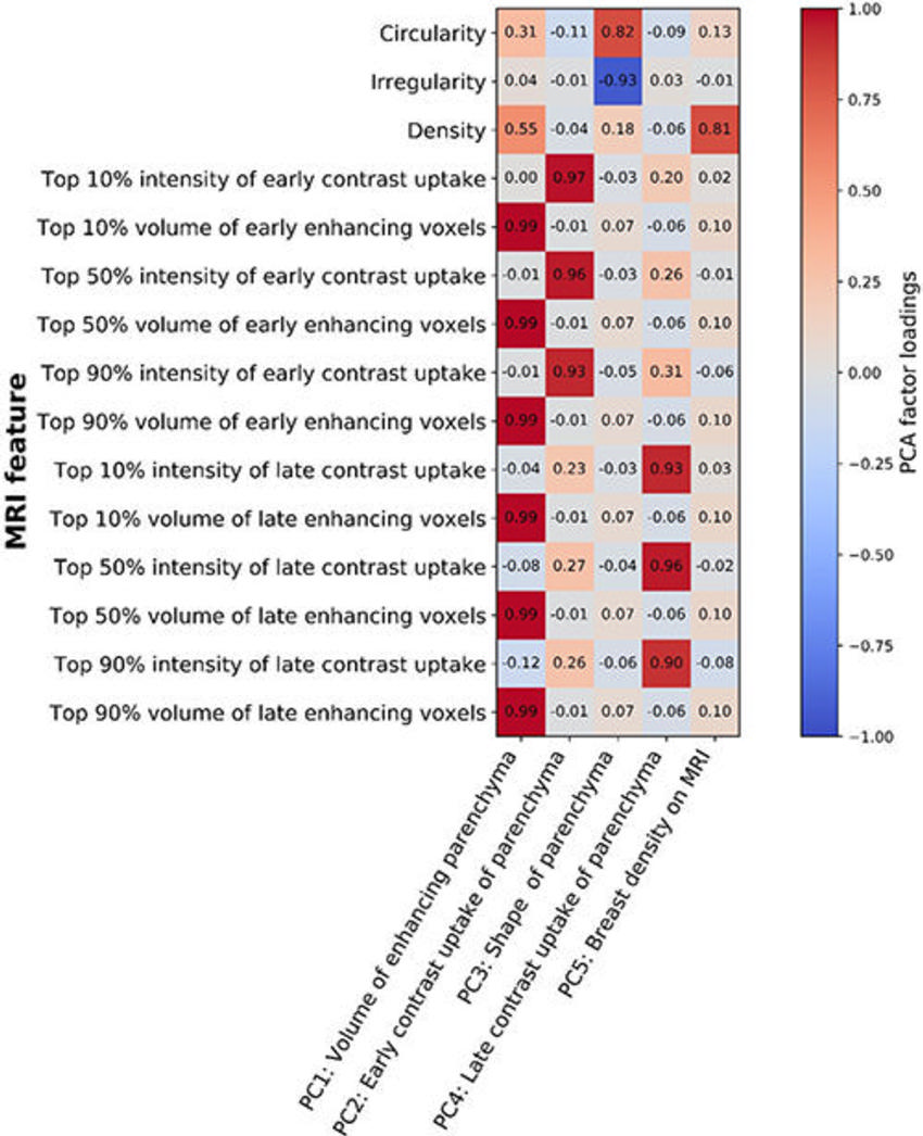
Figure 2. Heatmap shows the correlation between the 15 quantitative parenchyma features evaluated on baseline MRI scans and the principal components identified as accounting for 96% of the variance. The scale bar represents the principal component analysis (PCA) factor loading value, where the darkest red (value 1) represents a strong positive relationship and the darkest blue (value −1) represents a strong negative relationship.
High-res (TIF) version
(Right-click and Save As)
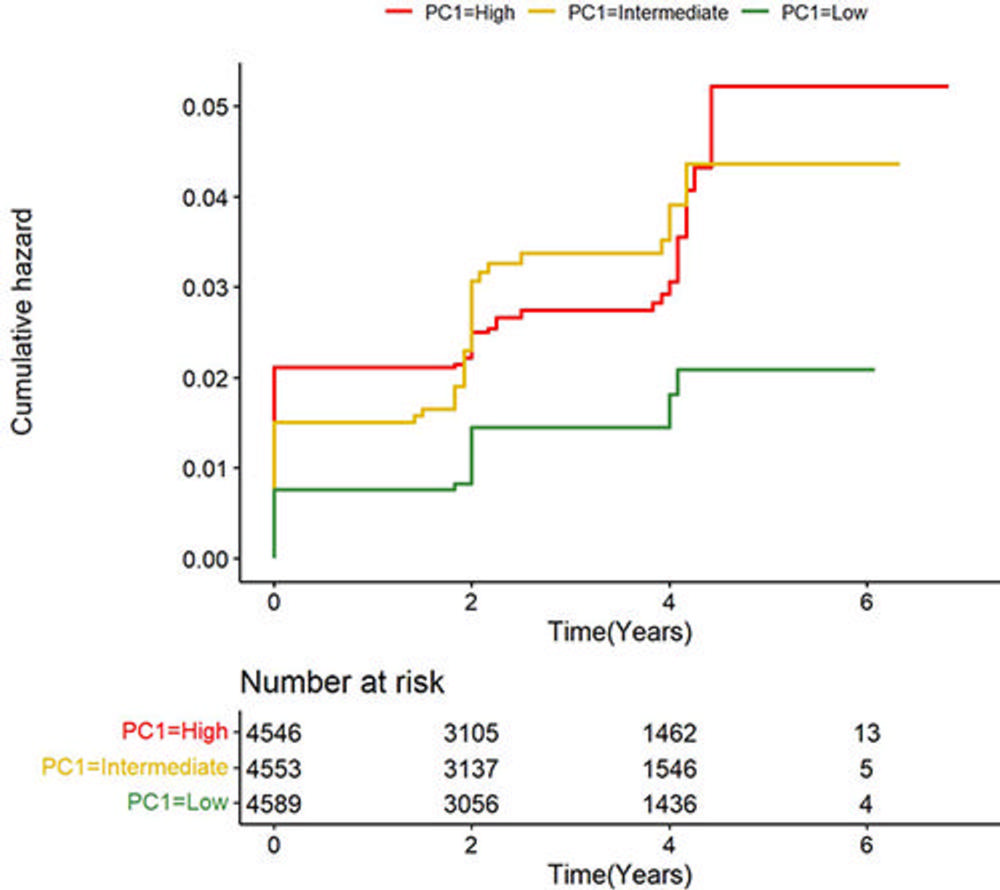
Figure 3. Kaplan-Meier curves for the occurrence of breast cancer stratified by tertiles of volume of enhancing parenchyma. Women in the high tertile (red line) and the intermediate tertile (yellow line) of volume of enhancing parenchyma had a higher cumulative hazard for breast cancer occurrence than those in the low tertile (green line) (log-rank test, P = .001 and P = .014, respectively). This analysis was adjusted for the clinical characteristics of age, body mass index, and background parenchymal enhancement category using inverse probability weighting. PC = principal component.
High-res (TIF) version
(Right-click and Save As)
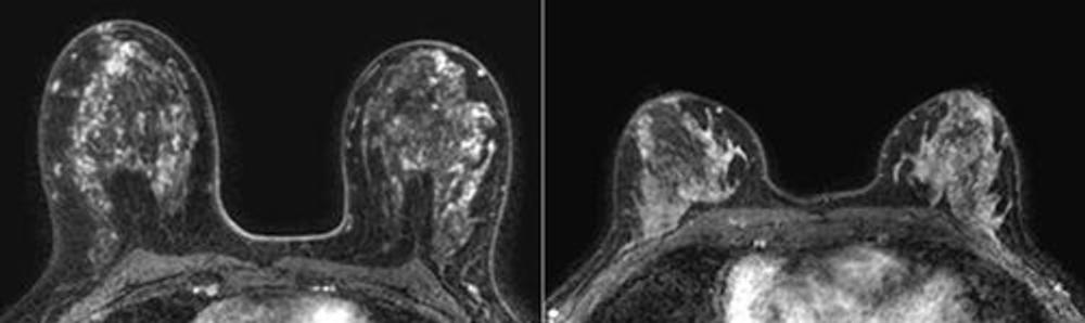
Figure 4. Representative transverse dynamic contrast-enhanced MRI scans in patients in the low and high tertiles of volume of enhancing parenchyma. Please note differences in high and low volumes of enhancing parenchyma are not visible to the naked eye on MRI scans. Left: Baseline MRI scan in a 50-year-old woman with a body mass index (BMI) of 23 and marked background parenchymal enhancement (BPE) who was stratified into the high tertile of volume of enhancing parenchyma. Cancer was detected with MRI in the second screening round. Right: Baseline MRI scan in a 51-year-old woman with a BMI of 24 and marked BPE who was stratified into the low tertile of volume of enhancing parenchyma. No cancer was detected during 6 years of follow-up.
High-res (TIF) version
(Right-click and Save As)