Photon-counting CT Noninvasively Detects Heart Disease in High-Risk Patients
Released: June 20, 2023
At A Glance
- Ultra-high-resolution photon-counting CT enables noninvasive detection of coronary artery disease in patients at high risk.
- Standard CT angiography is difficult in a high-risk population due to the high prevalence of calcifications and stents, which skew the results.
- UHR-CCTA is feasible for high-risk patients, in whom the benefits outweigh the risks, but should not be applied to all patients referred for cardiac CT imaging.
- RSNA Media Relations
1-630-590-7762
media@rsna.org - Linda Brooks
1-630-590-7738
lbrooks@rsna.org - Imani Harris
1-630-481-1009
iharris@rsna.org
OAK BROOK, Ill. — New ultra-high-resolution CT technology enables excellent image quality and accurate diagnosis of coronary artery disease in high-risk patients, a potentially significant benefit for people previously ineligible for noninvasive screening, according to a study published in Radiology, a journal of the Radiological Society of North America (RSNA).
Coronary artery disease is the most common form of heart disease. Coronary CT angiography (CCTA) is highly effective for ruling out coronary artery disease in patients at low or intermediate risk for the disease. Unfortunately, CCTA in a high-risk population is difficult due to a high prevalence of coronary calcifications and stents. Coronary calcifications tend to "bloom" on CCTA, making them appear more extensive than they really are. This results in overestimation of blockages and plaque and false-positive results.
"Consequently, patients may undergo unnecessary, often invasive, testing," said study lead author Muhammad T. Hagar, M.D., from the Department of Diagnostic and Interventional Radiology at the University of Freiburg in Freiburg, Germany. "This is the reason why current guidelines do not recommend using CCTA in high-risk individuals."
Ultra-high-resolution coronary CT angiography (UHR-CCTA) is a promising tool for the noninvasive assessment of patients at high risk for coronary artery disease. Because it uses recently introduced photon-counting CT scanners, it has not been extensively studied.
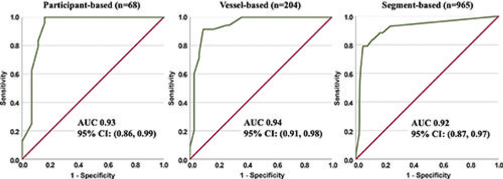
Dr. Hagar and colleagues compared the diagnostic accuracy of UHR-CCTA with that of the reference standard of invasive coronary angiography (ICA) in 68 patients. The patients had severe aortic valve stenosis, a common, serious valve disease that reduces or blocks blood flow from the heart to the aorta.

UHR-CCTA was highly sensitive and specific for coronary artery disease detection. It delivered a median overall image quality score of 1.5 on the 5-point Likert scale, where 1 is excellent and 5 is nondiagnostic. Almost 80% of segments rated as good or excellent.
The results suggest that the benefits of noninvasive imaging may soon be available to high-risk patients, Dr. Hagar said.
"It appears that the spectrum of patients benefiting from undergoing non-invasive CCTA has been significantly broadened by photon-counting detector technology," he said. "This is excellent news for these patients and the imaging community."
The high resolution of UHR-CCTA results from a greater number of emitted photons. This also increases radiation exposure compared with conventional CT scanners. However, Dr. Hagar noted that the technology is at a very early stage and that researchers are developing methods to reduce the amount of radiation exposure.
"Currently, the technique is feasible for high-risk patients, in whom the benefits outweigh the risks, but should not be applied to all patients referred for cardiac CT imaging," he said.
While photon-counting CT is relatively scarce worldwide, experts anticipate that the technology will become more prevalent in the next 10 years.
"At the University of Freiburg, we had the privilege to work with the technology since its introduction and I am convinced that photon-counting CT is the beginning of a new generation of CT scanners, similar to the introduction to multislice CT 30 years ago," Dr. Hagar said.
The researchers are exploring the diagnostic ability of photon-counting CT technology in other clinical scenarios such as oncological imaging. In the field of cardiac imaging, they are expanding their research to include subgroups for whom CT imaging is currently not feasible, such as patients with coronary stents. They also are looking into heart muscle assessment with photon-counting CT. Early findings suggest that the technology may improve soft tissue resolution. This would greatly benefit disease characterization.
"For me, these are exciting times, and it is just great to be part of a very active group working with this new technology," Dr. Hagar said.
"Accuracy of Ultrahigh-Resolution Photon-counting CT for Detecting Coronary Artery Disease in a High-Risk Population." Collaborating with Dr. Hagar were Martin Soschynski, M.D., Ruben Saffar, M.D., Alexander Rau, M.D., Jana Taron, M.D., Jakob Weiss, M.D., Thomas Stein, M.S., Sebastian Faby, Ph.D., Constantin von zur Muehlen, M.D., Philipp Ruile, M.D., Christopher L. Schlett, M.D., M.P.H., Fabian Bamberg, M.D., M.P.H., and Tobias Krauss, M.D.
In 2023, Radiology is celebrating its 100th anniversary with 12 centennial issues, highlighting Radiology's legacy of publishing exceptional and practical science to improve patient care.
Radiology is edited by Linda Moy, M.D., New York University, New York, N.Y., and owned and published by the Radiological Society of North America, Inc. (https://pubs.rsna.org/journal/radiology)
RSNA is an association of radiologists, radiation oncologists, medical physicists and related scientists promoting excellence in patient care and health care delivery through education, research and technologic innovation. The Society is based in Oak Brook, Illinois. (RSNA.org)
For patient-friendly information on CT angiography, visit RadiologyInfo.org.
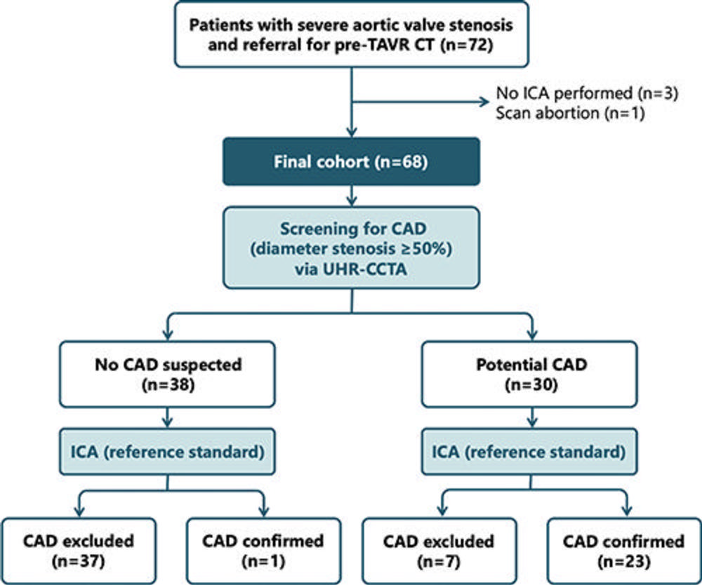
Figure 1. Flowchart of participant inclusion and exclusion criteria and coronary artery disease (CAD) diagnosis. CCTA = coronary CT angiography, ICA = invasive coronary angiography, TAVR = transcatheter aortic valve replacement, UHR = ultrahigh resolution.
High-res (TIF) version
(Right-click and Save As)
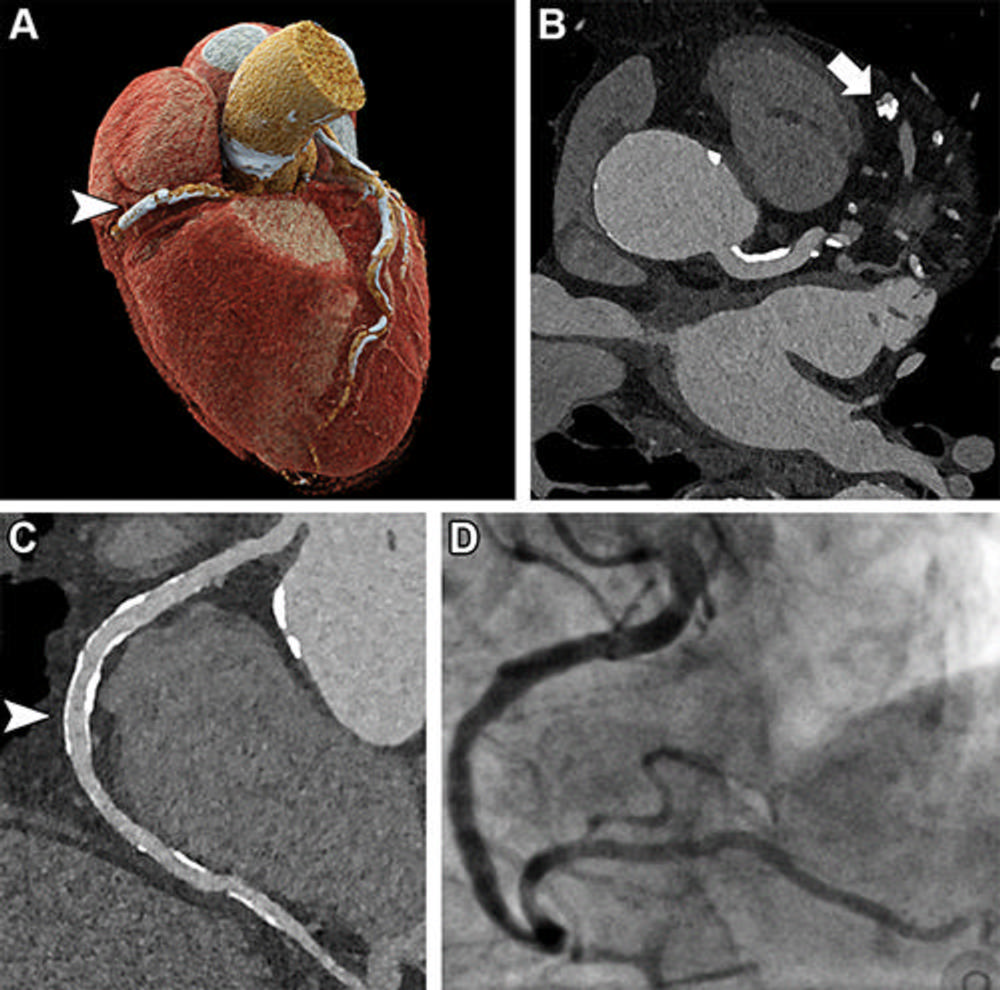
Figure 2. Ultrahigh-resolution (UHR) coronary CT angiography (CCTA) in an 85-year-old man before transcatheter aortic valve replacement. Despite a stent in the right coronary artery and very severe coronary sclerosis with an Agatston score of 4162, diagnostic visualization of the coronary arteries succeeded, and obstructive coronary artery disease was excluded on CT images. (A) Three-dimensional cinematic rendering of the heart. The stent (arrowhead) is visible in the middle segment of the right coronary artery. (B) UHR CCTA with 0.2-mm axial sections. The lumen (arrow) of the severely calcified distal left anterior descending artery can be assessed without artifacts. (C) Curved multiplanar reformations of the right coronary artery with a diagnostic display of the stent lumen (arrowhead). (D) Invasive coronary angiography enables exclusion of in-stent stenosis.
High-res (TIF) version
(Right-click and Save As)
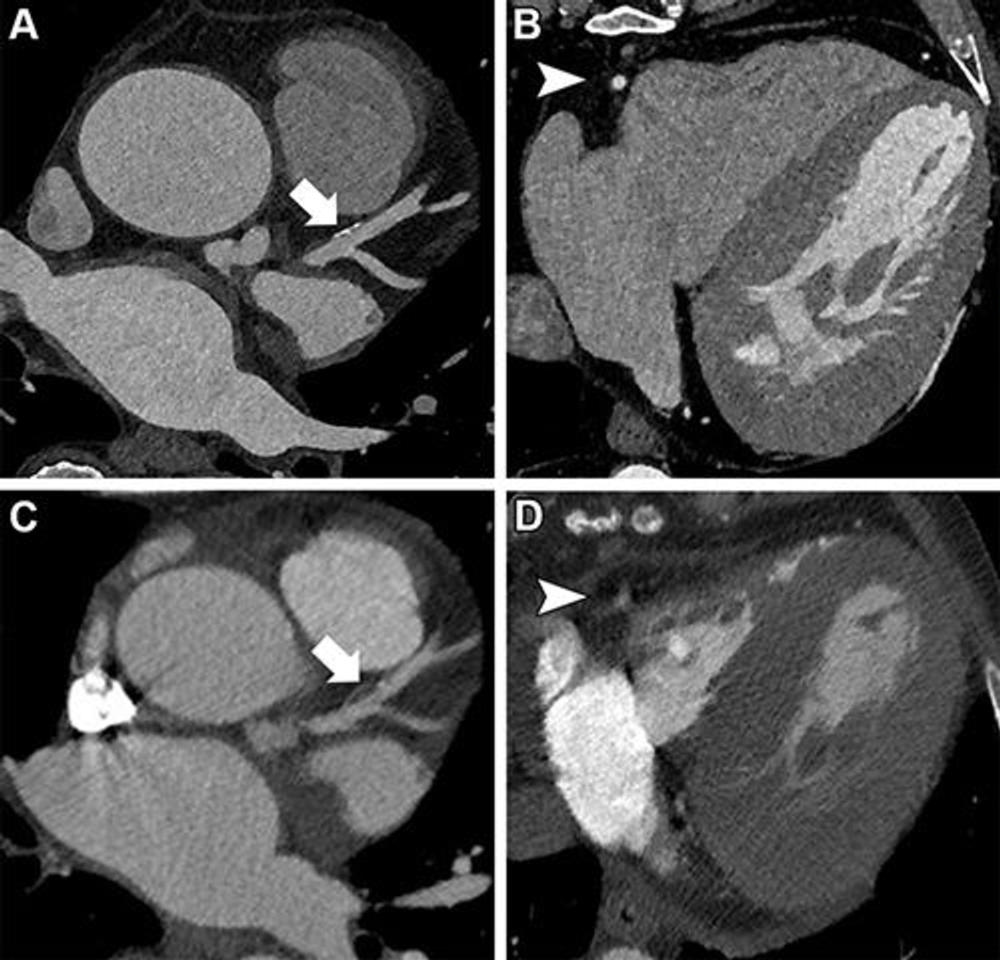
Figure 3. Images in a 67-year-old man with severe aortic valve stenosis recurrence after surgical aortic valve replacement 6 years ago and referral for valve-in-valve transcatheter aortic valve replacement CT. (A, B) Ultrahigh-resolution (UHR) coronary CT angiography (CCTA) on a first-generation dual-energy photon-counting scanner. (C, D) Cardiac CT images obtained with a second-generation dual-source energy-integrated CT scanner for planning this participant’s first aortic valve replacement 6 years prior to UHR CCTA. Note the superior image quality of UHR CCTA regarding vessel sharpness of the right coronary artery (arrowhead in B and D) and plaque visualization in the left anterior descending artery (arrow in A and C). Heart rates were comparable between UHR CCTA and cardiac CT (59 beats per minute vs 64 beats per minute, respectively), and contrast media and scan mode (electrocardiography-synchronized retrospective spiral scan) were similar.
High-res (TIF) version
(Right-click and Save As)