Study Examines Heart Inflammation after COVID Vaccine
Released: March 09, 2023
At A Glance
- Researchers found inflammation in the heart in only two patients with acute myocarditis when evaluated two months after COVID-19 vaccination.
- The cardiac PET/MRI study looked at 54 patients, including 17 with myocarditis.
- Patients without myocarditis had no heart inflammation or biomarker abnormalities at two-month follow-up, regardless of whether they reported symptoms.
- RSNA Media Relations
1-630-590-7762
media@rsna.org - Linda Brooks
1-630-590-7738
lbrooks@rsna.org - Imani Harris
1-630-481-1009
iharris@rsna.org
OAK BROOK, Ill. (March 9, 2023) — Researchers found evidence of heart muscle inflammation in a small number of patients with acute myocarditis after COVID-19 vaccination, but not in patients without acute myocarditis, according to a study published in Radiology: Cardiothoracic Imaging, a journal of the Radiological Society of North America (RSNA).

“To our knowledge, this is the first prospective study to report comprehensive cardiac investigations and imaging in both symptomatic and asymptomatic patients after COVID-19 vaccination,” said the study’s senior author Kate Hanneman, M.D., M.P.H., associate professor in the Department of Medical Imaging and director of cardiac imaging research at the University of Toronto in Toronto, Canada.
Billions of people worldwide have received at least one dose of a COVID-19 vaccine. Some instances of myocarditis (non-ischemic inflammation of the heart muscle) have been reported following administration of mRNA-based COVID-19 vaccines. Myocarditis can affect the heart’s rhythm and ability to pump blood and may leave behind lasting damage in the form of scarring of the heart muscle.
Most instances of myocarditis after COVID-19 vaccination have occurred in adolescent and young adult males. However, the overall risk is very low.
Some patients may experience cardiac symptoms after vaccination, including shortness of breath, palpitations, and chest pain, yet do not meet diagnostic criteria for acute myocarditis.
“We know that some patients are at risk of myocarditis following mRNA-based COVID-19,” Dr. Hanneman said. “However, there are limited data on potential myocardial tissue changes in patients who are asymptomatic after COVID-19 vaccination. Similarly, we have very limited data to date on patients who present with new symptoms after vaccination but do not meet diagnostic criteria for acute myocarditis.”
Cardiac MRI plays an important role in the assessment of acute myocarditis with unparalleled ability for noninvasive characterization of myocardial tissue. Cardiac fluorine 18 (18F) fluorodeoxyglucose (FDG) PET allows for assessment of changes in myocardial metabolism.
For this prospective study, Dr. Hanneman and a multi-disciplinary team of radiology, cardiology and vascular biology researchers set out to investigate cardiac effects of COVID-19 vaccination at two-month follow-up and relate cardiac symptoms to myocardial tissue changes on FDG PET/MRI, blood biomarkers, health-related quality of life and adverse outcomes.
Fifty-four participants were evaluated a median of 72 days after COVID-19 vaccination. Seventeen were symptomatic with myocarditis, 17 were symptomatic without myocarditis, and 20 were asymptomatic.
No participants in the symptomatic without myocarditis or asymptomatic groups had focal FDG uptake, myocardial edema or impaired ventricular function. Two participants with symptomatic myocarditis had focal FDG uptake. Health-related quality of life was lower in the symptomatic myocarditis group than the asymptomatic group. There were no adverse cardiac events beyond myocarditis in any participant.
“None of the symptomatic participants in our study who did not meet diagnostic criteria for acute myocarditis had elevated troponin levels, impaired left ventricular function, or detectable cardiac inflammation two months after COVID-19 vaccination,” Dr. Hanneman said. “This suggests that symptoms alone are a poor indicator of myocardial injury after vaccination.”
The results also suggested that subclinical myocardial injury is not common after COVID-19 vaccination, based on normal cardiac PET, ECG, and blood biomarker findings in the asymptomatic patient group. The researchers hope these findings reassure patients who did not experience symptoms after COVID-19 vaccination but worry about the possibility of subclinical cardiac disease.
Dr. Hanneman cautioned that further study is needed to investigate non-cardiovascular causes of symptoms after vaccination in individuals who do not meet diagnostic criteria for acute myocarditis.
“Myocardial Inflammation on FDG PET/MRI and Clinical Outcomes in Symptomatic and Asymptomatic Participants after COVID-19 Vaccination.” Collaborating with Dr. Hanneman were Constantin Arndt Marschner, M.D., Paaladinesh Thavendiranathan, M.D., S.M., Dakota Gustafson, B.Sc., Kathryn L. Howe M.D., Ph.D., Jason E. Fish, Ph.D., Robert M. Iwanochko, M.D., Rachel M. Wald, M.D., Husam Abdel-Qadir, M.D., Slava Epelman, M.D., Ph.D., Angela M. Cheung, M.D., Ph.D., and Rachel Hong, B.Sc.
Radiology: Cardiothoracic Imaging is edited by Suhny Abbara, M.D., University of Texas Southwestern Medical Center, Dallas, and owned and published by the Radiological Society of North America, Inc.(https://pubs.rsna.org/journal/cardiothoracic)
RSNA is an association of radiologists, radiation oncologists, medical physicists and related scientists promoting excellence in patient care and health care delivery through education, research, and technologic innovation. The Society is based in Oak Brook, Illinois. (RSNA.org)
For patient-friendly information on cardiac MRI, visit RadiologyInfo.org.
Press Resources:
Video (MP4):

Video 1. Dr. Kate Hanneman discusses her research on heart inflammation after COVID vaccine.
Download MP4
(Right-click and Save As)
Images (JPG, TIF):
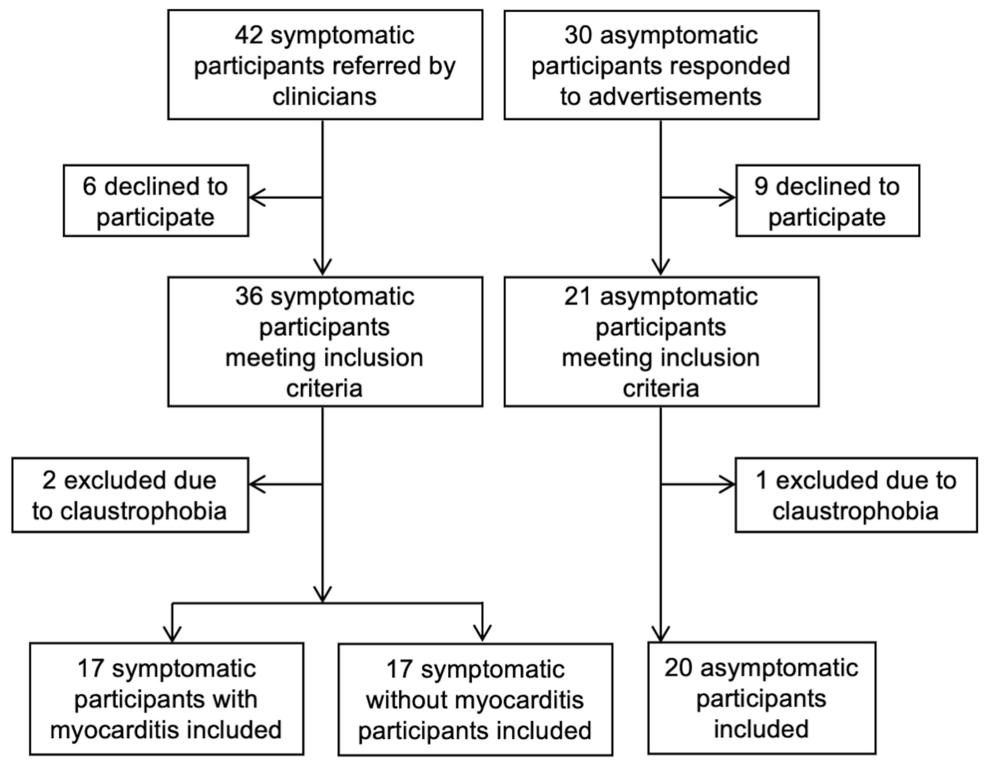
Figure 1. Flowchart detailing participant selection.
High-res (TIF) version
(Right-click and Save As)
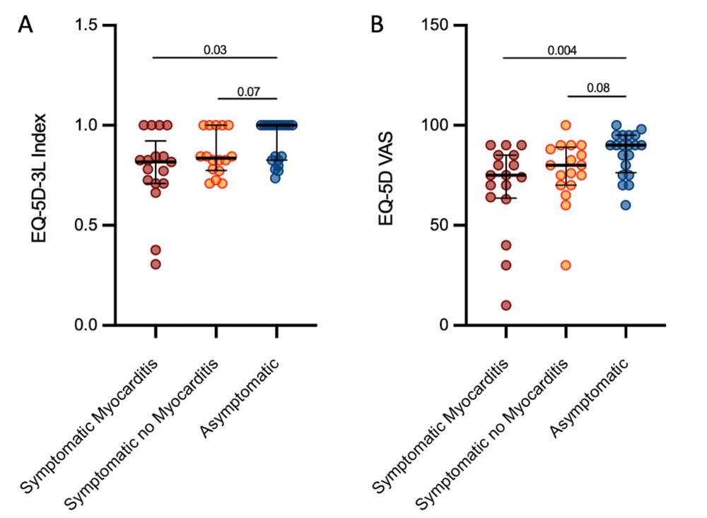
Figure 2. Quality of Life at Short-term Follow-up by Participant Group. Scatterplots for (A) EQ-5D-3L index and (B) EQ-5D-3L visual analogue scale (VAS) depict individual patient data points with error bars displayed as median and interquartile range. P-values are for comparisons between groups as indicated.
High-res (TIF) version
(Right-click and Save As)
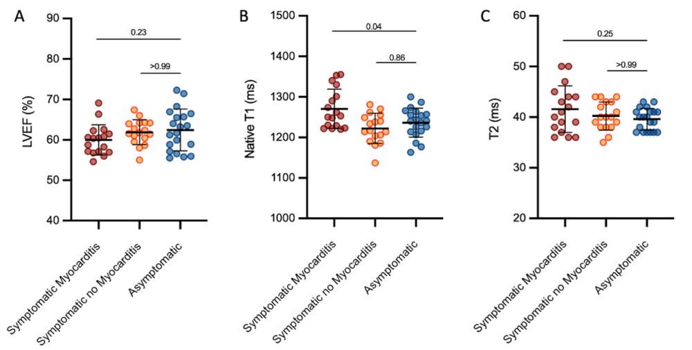
Figure 3. Cardiac PET/MRI findings at Follow-up by Participant Group. Scatterplots for continuous PET/MRI parameters of (A) left ventricular ejection fraction [LVEF], (B) native T1 mapping, and (C) native T2 mapping depict individual patient data points with error bars displayed as mean and standard deviation. P-values are for comparisons between groups as indicated.
High-res (TIF) version
(Right-click and Save As)
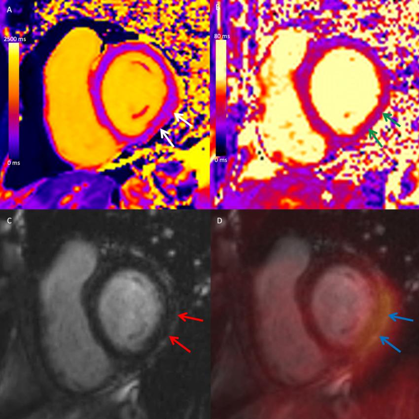
Figure 4. Cardiac PET/MRI in a Participant with Myocarditis after COVID-19 Vaccination. Combined cardiac fluorine 18 fluorodeoxyglucose (FDG) PET/MRI in a symptomatic female participant between 41-50 years of age two months after a diagnosis of myocarditis following COVID-19 vaccination. Short-axis mid-ventricular native (A) T1 and (B) T2 maps demonstrate high T1 and T2 values in the subepicardial inferior and inferolateral wall (white and green arrows, respectively). (C) Short-axis late gadolinium enhancement (LGE) image demonstrates corresponding subepicardial LGE (red arrows). (D) Fused LGE and FDG PET image demonstrates corresponding focal FDG uptake (blue arrows), in keeping with myocardial inflammation.
High-res (TIF) version
(Right-click and Save As)