Thrombectomy Comparable to Medical Management for Strokes
Released: February 14, 2023
At A Glance
- A surgical procedure called thrombectomy is a safe and feasible treatment option for a rare type of stroke.
- For the study, researchers compared thrombectomy with drug treatment in 154 patients and assessed early outcome within the first 24 hours after treatment.
- Within the first 24 hours after treatment, thrombectomy patients had similar outcomes to those who received medications to dissolve the clot.
- RSNA Media Relations
1-630-590-7762
media@rsna.org - Linda Brooks
1-630-590-7738
lbrooks@rsna.org - Imani Harris
1-630-481-1009
iharris@rsna.org
OAK BROOK, Ill. — A surgical procedure called thrombectomy is a safe and feasible treatment option for a rare type of stroke, according to a study published in Radiology, a journal of the Radiological Society of North America (RSNA).
Thrombectomy is commonly performed to remove blood clots from brain arteries. It is often done to treat strokes arising from blood clots in the larger arteries that supply blood to the brain. When done promptly, thrombectomy can sometimes be more successful than clot-busting drugs in mitigating long-term damage to the brain.
Recent advances in technology have made it possible to perform thrombectomy in some of the narrower vessels in the head—vessels like the anterior cerebral arteries that arise from the internal carotid artery. However, evidence supporting a potential benefit of thrombectomy for these vessels remains unknown.
As part of the Treatment fOr Primary Medium vessel Occlusion Stroke, or TOPMOST trial, researchers in Germany compared thrombectomy with medical treatment in 154 patients. The patients all had primary isolated anterior cerebral artery medium vessel occlusions, or obstructions. The patients underwent thrombectomy or best medical treatment, which typically involves medications to dissolve the clot and reduce blood pressure. In some cases, patients may receive intravenous thrombolysis, the introduction of clot-busting drugs to the bloodstream or directly to the site of clot.
The researchers assessed early outcome, or outcome within the first 24 hours after treatment, and longer-term functional outcome. They also looked at safety with a focus on bleeding in the brain and death.
The results showed that thrombectomy was a safe and technically feasible option. Within the first 24 hours after treatment, thrombectomy patients had similar outcomes to those who received best medical treatment alone with or without intravenous thrombolysis. Longer term, both groups had similar clinical and functional outcomes. Mortality rates were similar in both groups.
“Based on our study, both treatment options appear to be effective and safe,” said study lead author Lukas Meyer, M.D., from the Department of Diagnostic and Interventional Neuroradiology at University Medical Center Hamburg-Eppendorf in Hamburg, Germany. “The overall results of the study are consistent with a growing body of literature suggesting that thrombectomy may have a role in the treatment of this type of stroke. Eligible patients should therefore be randomized to ongoing prospective trials whenever possible.”
Dr. Meyer emphasized that the type of blockages his team studied are rare, and all the centers involved in this study were tertiary stroke centers with a high level of expertise in these kinds of interventions.
Selection of patients for thrombectomy remains a key issue, Dr. Meyer said, especially when patients are not eligible for randomization in a designated trial.
“As we anticipate an increasing number of patients with medium vessel occlusions being treated with thrombectomy, age and eloquence of symptoms in particular are factors that should be considered when making treatment decisions,” he said. “Ultimately, the results of ongoing randomized trials will provide further insight into this subgroup.”
Dr. Meyer plans to conduct further analysis of a subgroup of these patients to gain a deeper understanding of procedural and clinical outcomes. He also wants to identify potential surrogate markers that can predict clinical course.
“By focusing on this specific patient population, we hope to further advance our knowledge and improve patient outcomes,” he said.
“Thrombectomy versus Medical Management for Isolated Anterior Cerebral Artery Stroke: An International Multicenter Registry Study.” Collaborating with Dr. Meyer were Paul Stracke, M.D., Gabriel Broocks, M.D., Mohamed Elsharkawy, M.D., Peter Sporns, M.D., Eike I. Piechowiak, M.D., Johannes Kaesmacher, M.D., Christian Maegerlein, M.D., Moritz Roman Hernandez Petzsche, M.D., Hanna Zimmermann, M.D., Weis Naziri, M.D., Nuran Abdullayev, M.D., Christoph Kabbasch, M.D., Elie Diamandis, M.D., Maximilian Thormann, M.D., Volker Maus, M.D., Sebastian Fischer, M.D., Markus Möhlenbruch, M.D., Charlotte S. Weyland, M.D., Marielle Ernst, M.D., Ala Jamous, M.D., Dan Meila, M.D., Milena Miszczuk, M.D., Eberhard Siebert, M.D., Stephan Lowens, M.D., Lars Udo Krause, M.D., Leonard Yeo, M.B.B.S., Benjamin Tan, M.B.B.S., Anil Gopinathan, M.D., Juan F. Arenillas-Lara, M.D., Pedro Navia, M.D., Eytan Raz, M.D., Maksim Shapiro, M.D., Fabian Arnberg, M.D., Kamil Zeleňák, M.D., Mario Martinez-Galdamez, M.D., Maria Alexandrou, M.D., Andreas Kastrup, M.D., Panagiotis Papanagiotou, M.D., André Kemmling, M.D., Franziska Dorn, M.D., Marios Psychogios, M.D., Tommy Andersson, M.D., René Chapot, M.D., Jens Fiehler, M.D., and Uta Hanning, M.D., for the TOPMOST Study Group.
In 2023, Radiology is celebrating its 100th anniversary with 12 centennial issues, highlighting Radiology’s legacy of publishing exceptional and practical science to improve patient care.
Radiology is edited by Linda Moy, M.D., New York University, New York, N.Y., and owned and published by the Radiological Society of North America, Inc. (https://pubs.rsna.org/journal/radiology)
RSNA is an association of radiologists, radiation oncologists, medical physicists and related scientists promoting excellence in patient care and health care delivery through education, research, and technologic innovation. The Society is based in Oak Brook, Illinois. (RSNA.org)
For patient-friendly information on stroke, visit RadiologyInfo.org.
Images (JPG, TIF):

Figure 1. Flowchart of patient inclusion before and after propensity score matching (PSM). ACA = anterior cerebral artery, BMT = best medical treatment, DMVO = distal medium vessel occlusion, MT = mechanical thrombectomy.
High-res (TIF) version
(Right-click and Save As)

Figure 2. (A–C) Axial perfusion CT (time-to-maximum, < seconds) at admission shows a bilateral deficit (arrows in A) in the territory of the anterior cerebral artery (ACA; ie, azygos variant) because of a distal occlusion of the A3 segment (white arrow, sagittal view in B). Stent retriever thrombectomy (black arrow, sagital view in B) was performed with full reperfusion shown on the final digital subtraction angiography image (arrow, sagittal view in C). (D) Follow-up at 24 hours shows no sign of infarction in the ACA on the axial contrast-unenhanced CT image.
High-res (TIF) version
(Right-click and Save As)

Figure 3. (A) Axial contrast-unenhanced CT image at admission shows no sign of early ischemic changes. (B) Digital subtraction angiography shows a proximal occlusion of the A3 segment (arrow, sagittal view). (C) After two thrombectomy attempts the affected territory underwent partial reperfusion (black arrow, sagittal view) with a distal remaining A5 occlusion (white arrow, sagittal view). (D) Follow-up with MRI at 24 hours shows signs of infarction in the distal territory of the anterior cerebral artery on axial diffusion-weighted image (arrow).
High-res (TIF) version
(Right-click and Save As)

Figure 4. Procedural endovascular complications and angiographic outcome stratified by occluded vessel segments. * Includes one case with an A4 occlusion. ACoA = anterior communicating artery, ENT = emboli to new territory, HI = hemorrhagic transformation, IP = itrogenic perforation, IVH = intraventricular hemorrhage, mTICI = modified thrombolysis in cerebral infarction, PH = parenchymal hemorrhage, SAH = subarachnoid hemorrhage, sICH = symptomatic intracerebral hemorrhage.
High-res (TIF) version
(Right-click and Save As)

Figure 5. Distribution of modified Rankin scale (mRS) scores at 90 days shows functional long-term outcome compared by treatment cohorts of best medical treatment (BMT) and endovascular treatment before and after propensity score matching (PSM). MT= mechanical thrombectomy.
High-res (TIF) version
(Right-click and Save As)

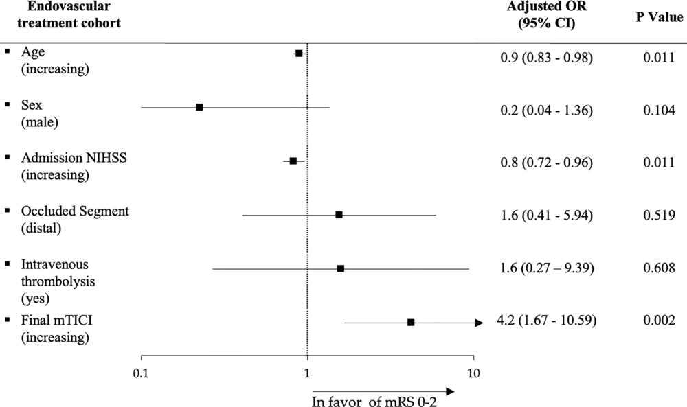
Figure 6. Multivariable logistic regression analysis for favorable functional outcome shows factors associated with modified Rankin scale (mRS) scores of 0–2 at 90-day follow-up in the endovascular treatment cohort. mTICI = modified thrombolysis in cerebral infarction, NIHSS = National Institutes of Health Stroke Scale, OR = odds ratio.
High-res (TIF) version
(Right-click and Save As)
