Photon-Counting CT Enables Lower Contrast Media for Aortic Imaging
Released: January 26, 2023
At A Glance
- Photon-counting detector CT is a new technology that can acquire high-quality images at lower contrast media volume than conventional CT.
- The study included 100 patients who underwent CT angiography with photon-counting CT of the aorta in the chest and abdomen.
- While contrast media used in these exams is considered very safe, there is a higher risk of adverse reactions in patients with kidney disease.
- RSNA Media Relations
1-630-590-7762
media@rsna.org - Linda Brooks
1-630-590-7738
lbrooks@rsna.org - Imani Harris
1-630-481-1009
iharris@rsna.org
OAK BROOK, Ill. (January 26, 2023) — Photon-counting detector CT reduces the amount of contrast needed for CT angiography (CTA) while maintaining image quality, according to a new study published in Radiology: Cardiothoracic Imaging, a journal of the Radiological Society of North America (RSNA).
The portion of the aorta that passes through the chest and abdomen is prone to aneurysms. Risk increases with age. Doctors use CTA with contrast media to examine this region of the artery. While the iodinated contrast media used in these exams is considered very safe, there is a higher risk of adverse reactions in patients with kidney disease, or nephropathy.
"Many patients undergoing CTA of the aorta are elderly, and some may suffer from a certain degree of nephropathy," said senior author Hatem Alkadhi, M.D., M.P.H., from the Department of Diagnostic and Interventional Radiology at University Hospital Zurich in Zurich, Switzerland. "Thus, they might be at risk for a further reduction of their kidney function due to contrast media administration."
Using less contrast media is also good for the environment, as iodine breakdown products have harmful environmental effects. In addition, the supply of contrast media has suffered from shortages due to COVID-19-related disruptions in the pharmaceutical supply chain.

Photon-counting detector CT is a new technology that can acquire high-quality images at lower contrast media volume than conventional CT scanners that rely on energy-integrating detectors. It also offers diagnostic quality images at lower radiation dose than those of conventional CT.
Dr. Alkadhi and his University Hospital colleague Kai Higashigaito, M.D., led a study that looked at a low-volume contrast media protocol with photon-counting detector CT for CTA of the aorta in the chest and abdomen.
The study included 100 people, mean age 75. Patients underwent CTA with photon-counting detector CT of the aorta in the chest and abdomen. The patients had received a previous CTA with conventional CT at equal radiation doses.
Photon-counting CT had a higher contrast-to-noise ratio, a key measure of image quality, which translates to a low-volume contrast media protocol. Contrast media volume was reduced by 25%.
Two independent readers rated the image quality of photon-counting CT as higher than that of conventional CT at an equal radiation dose.
"We showed that the improved image quality of CTA with photon-counting detector CT systems can be used to reduce the amount of administered contrast media to the patients, without reducing the diagnostic yield of the examination," Dr. Alkadhi said. "Image quality remained at the same level as that of previous CT angiography examinations in the same patients using a conventional CT, despite the fact that we reduced the contrast media volume."
Based on the positive results of the study, Dr. Alkadhi and his colleagues have changed their clinical routine. They now adjust the contrast media protocols in patients undergoing CTA of the aorta in the chest and abdomen.
Besides its superior image quality and spatial resolution, photon-counting CT can provide detailed studies of tissue through its spectral imaging capability. Researchers are looking at this as a way to improve studies of the coronary arteries and the heart muscle.
"Based on the initial experience with this new exciting technology, I envision that this detector type will become the standard in CT scanners in the near future," Dr. Alkadhi said.
"CT Angiography of the Aorta Using Photon-counting Detector CT with Reduced Contrast Media Volume." Collaborating with Drs. Alkadhi and Higashigaito were Victor Mergen, M.D., Matthias Eberhard, M.D., E.B.C.R., Lisa Jungblut, M.D., Monika Hebeisen, M.Sc., Susan Rätzer, Bettina Zanini, Adrian Kobe, M.D., Katharina Martini, M.D., and André Euler, M.D.
Radiology: Cardiothoracic Imaging is edited by Suhny Abbara, M.D., University of Texas Southwestern Medical Center, Dallas, and owned and published by the Radiological Society of North America, Inc. (https://pubs.rsna.org/journal/cardiothoracic)
RSNA is an association of radiologists, radiation oncologists, medical physicists and related scientists promoting excellence in patient care and health care delivery through education, research, and technologic innovation. The Society is based in Oak Brook, Illinois. (RSNA.org)
For patient-friendly information on CT angiography, visit RadiologyInfo.org.
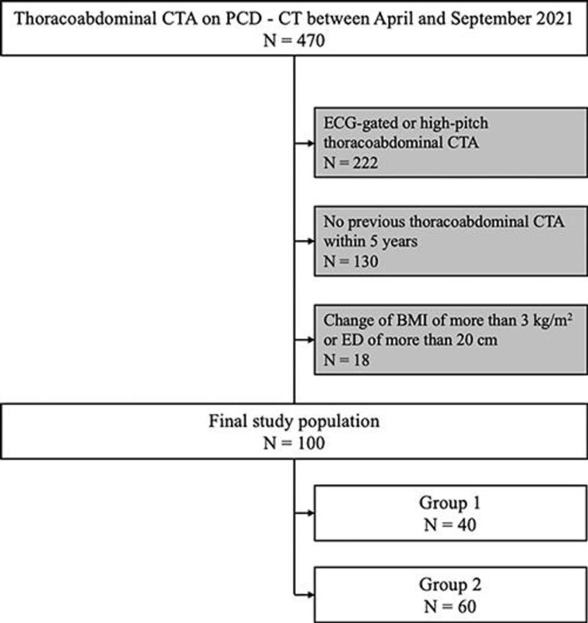
Figure 1. Study flowchart of participant selection based on inclusion and exclusion (dark gray) criteria. BMI = body mass index, CTA = CT angiography, ECG = electrocardiography, ED = effective diameter, PCD = photon-counting detector.
High-res (TIF) version
(Right-click and Save As)
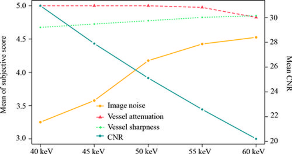
Figure 2. Contrast-to-noise ratio (CNR) and subjective image quality scores of photon-counting detector CT examinations for group 1 at each kiloelectron volt level. Note the diverging trends in objective and subjective image quality, with the highest objective and lowest subjective image quality at 40 keV and the lowest objective and highest subjective image quality at 60 keV. Scores are based on a five-point Likert scale: 5 = excellent, 4 = good, 3 = moderate, 2 = poor, 1 = nondiagnostic.
High-res (TIF) version
(Right-click and Save As)
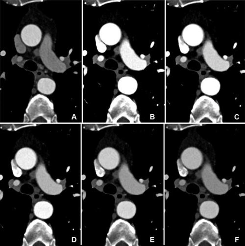
Figure 3. Comparison of image quality between EID CT and PCD CT using identical contrast media protocol and matched radiation dose. Transverse CTA images show the thoracic aorta at the level of the pulmonary trunk in a 62-year-old man in group 1, shown using identical window settings (window center: 350 HU, window width: 1000 HU). (A) Images from scans performed with a third-generation dual-source CT system with EIDs. (B–F) Images from PCD CT with VMI at energy levels from 40 to 60 keV. Time interval between scans was 11 months. Both scans were performed with the same contrast media protocol (volume, 70 mL). Note the higher vessel attenuation at lower energy levels for PCD CT. CTA = CT angiography, EID = energy-integrating detector, PCD = photon-counting detector, VMI = virtual monoenergetic images.
High-res (TIF) version
(Right-click and Save As)
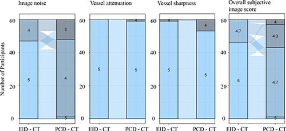
Figure 4. Subjective image quality scores of reader 1 for group 2. Alluvial diagram with vertical stacked bar charts of the subjective image scores from EID CT with standard contrast media volume and PCD CT at 50 keV VMI with reduced contrast media volume are shown. The blue and gray curved lines between columns represent the image score change for each individual participant. Interpretation of scores: 5 = excellent, 4 = good, 3 = moderate. EID = energy-integrating detector, PCD = photon-counting detector, VMI = virtual monoenergetic images.
High-res (TIF) version
(Right-click and Save As)
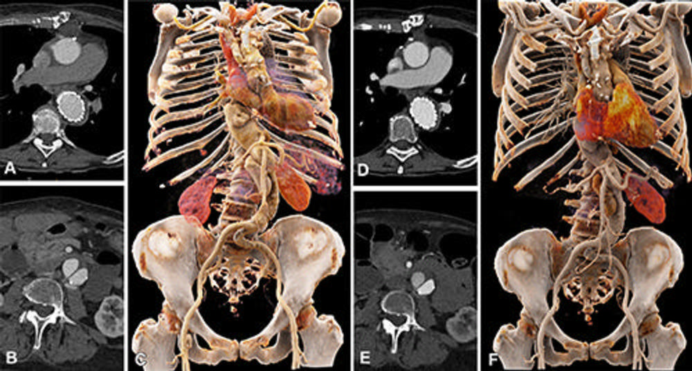
Figure 5. Comparison of image quality between EID CT with standard contrast media protocol and PCD CT with low-volume contrast media protocol using a matched radiation dose. Transverse and three-dimensional cinematic rendered images from thoracoabdominal CTA in a 71-year-old woman in group 2 are shown. (A–C) Images from third-generation EID CT with automated tube voltage selection of 90 kVp. (D–F) Images from PCD CT with reduced contrast media volume of 52.5 mL and VMI at 50 keV. Time interval between scans was 6 months. CTA = CT angiography, EID = energy-integrating detector, PCD = photon-counting detector, VMI = virtual monoenergetic images.
High-res (TIF) version
(Right-click and Save As)