Lasting Lung Damage Seen in Children and Teens after COVID
Released: September 20, 2022
At A Glance
- Children and adolescents who have recovered from COVID-19 or have long COVID show persistent lung damage and dysfunction.
- MRI allowed the researchers to derive the V/Q match, a measure of air and blood flow in the lungs.
- The V/Q match was 62% in the recovered group and 60% in the long COVID group—both considerably lower than the 81% match in healthy controls.
- RSNA Media Relations
1-630-590-7762
media@rsna.org - Linda Brooks
1-630-590-7738
lbrooks@rsna.org - Imani Harris
1-630-481-1009
iharris@rsna.org
OAK BROOK, Ill. (September 20, 2022) — Children and adolescents who have either recovered from COVID-19 or have long COVID show persistent lung damage on MRI, according to a study published in Radiology, a journal of the Radiological Society of North America (RSNA).

COVID-19 is an infectious disease caused by the SARS-CoV-2 virus. Since emerging in late 2019, it has killed more than 5 million people worldwide. The lungs are the primary target for the virus.
Study of the disease’s long-term effects has accelerated as the number of COVID survivors climbs and more people are diagnosed with long COVID. The World Health Organization defines long COVID as involving symptoms that persist for a minimum of 12 weeks and other factors, such as symptoms that result in a new health limitation or worsening of a pre-existing underlying medical condition.
The nature of the post-acute phase of the infection is poorly understood in younger people. CT has shown persistent damage to the lungs in adults, but CT uses ionizing radiation and has limited diagnostic value in children, where lung changes due to COVID-19 are less pronounced.
“We conceived this study when the evidence for long- or post-COVID cases in adults was growing,” said study senior author Ferdinand Knieling, M.D., specialist in pediatrics and adolescent medicine from the departments of Pediatrics and Adolescent Medicine at University Hospital Erlangen in Erlangen, Germany. “This was also when the first patients with unspecific symptoms were seen in our department, and parents started to ask about an association with a prior infection.”
Dr. Knieling and colleagues studied COVID-19’s effects in children and adolescents using low-field MRI. The technology relies on a lower magnetic field than conventional MRI and allows for free breathing, meaning the subjects do not have to hold their breath during imaging. This makes scanning more feasible in children.
“As parents, we also wanted to find what risks an infection might have,” Dr. Knieling said. “Luckily, our departments teamed up to use their brand-new MRI scanner designed for investigations in children and adolescents.”
The researchers looked at changes in lung structure and function in 54 children and adolescents (mean age 11 years) with previous SARS-CoV-2 infection. Of the 54 patients, 29 had recovered, and 25 had long COVID. All but one of the patients had been unvaccinated at the time of original infection.
None of the COVID-19 group required hospital admission during the primary infection period. Shortness of breath, impaired attention, headache, fatigue and loss of smell were the most commonly reported symptoms at the time of the study. Results from the COVID-19 group were compared with those from nine healthy controls.
MRI allowed the researchers to derive the V/Q match, a measure of air and blood flow in the lungs. If lungs are working properly, the air and blood flow should match.
V/Q matches showed persistent pulmonary dysfunction in the patients who had recovered from COVID-19 and in those with long COVID. The V/Q match was 62% in the recovered group and 60% in the long COVID group—both considerably lower than the 81% match in healthy controls.
“Persistent symptoms after COVID still cause diagnostic odysseys, and this is especially true for young people,” Dr. Knieling said. “Our findings illustrate that caring for these patients is a multidisciplinary challenge.”
Long-term implications of these lung changes remain unclear, but the results warrant further surveillance of persistent lung damage in children and adolescents after COVID-19, Dr. Knieling said. Lung MRI is already widely available, he noted, making these imaging approaches easy to integrate into clinical routine care. More research will help show the full potential of MRI in COVID-19 survivors.
“A follow-up trial has already started, and we seek to understand how findings change over time,” Dr. Knieling said. “Additionally, we will take closer looks at other organs to see how this correlates with our findings.”
“Pulmonary Dysfunction after Pediatric COVID-19.” Collaborating with Dr. Knieling were Rafael Heiss, M.D., Lina Tan, Sandy Schmidt, Adrian P. Regensburger, M.D., Franziska Ewert, M.D., Dilbar Mammadova, M.D., Adrian Buehler, M.Sc., Jens Vogel-Claussen, M.D., Andreas Voskrebenzev, Ph.D., Manfred Rauh, Ph.D., Oliver Rompel, M.D., Armin M. Nagel, M.D., Simon Lévy, M.D., Sebastian Bickelhaupt, M.D., Matthias S. May, M.D., Michael Uder, M.D., Markus Metzler, M.D., Regina Trollmann, M.D., Joachim Woelfle, M.D., and Alexandra L. Wagner, M.D.
Radiology is edited by David A. Bluemke, M.D., Ph.D., University of Wisconsin School of Medicine and Public Health, Madison, Wisconsin, and owned and published by the Radiological Society of North America, Inc. (https://pubs.rsna.org/journal/radiology)
RSNA is an association of radiologists, radiation oncologists, medical physicists and related scientists promoting excellence in patient care and health care delivery through education, research and technologic innovation. The Society is based in Oak Brook, Illinois. (RSNA.org)
For patient-friendly information on pediatric MRI, visit RadiologyInfo.org.
Images (JPG, TIF):
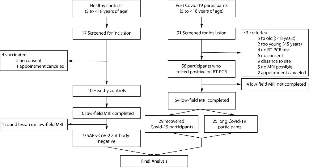
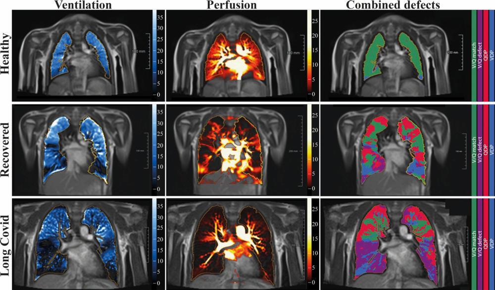
Figure 2. Free-breathing phase-resolved functional lung (PREFUL) low-field MRI at 0.55T with calculated parameters at an axial plane after automatic registration to a mid-expiration position and lung parenchyma segmentation. From left to right, representative color-coded images of functional show ventilation defects (VDP, blue), perfusion defects (QDP, red), ventilation/perfusion (V/Q match, green), ventilation/perfusion defects (V/Q defect, purple) in a healthy control (upper row, 7-year-old male), a participant recovered from COVID-19 (middle row, 10-year-old male) and a participant with long COVID (15-year-old male).
High-res (TIF) version
(Right-click and Save As)
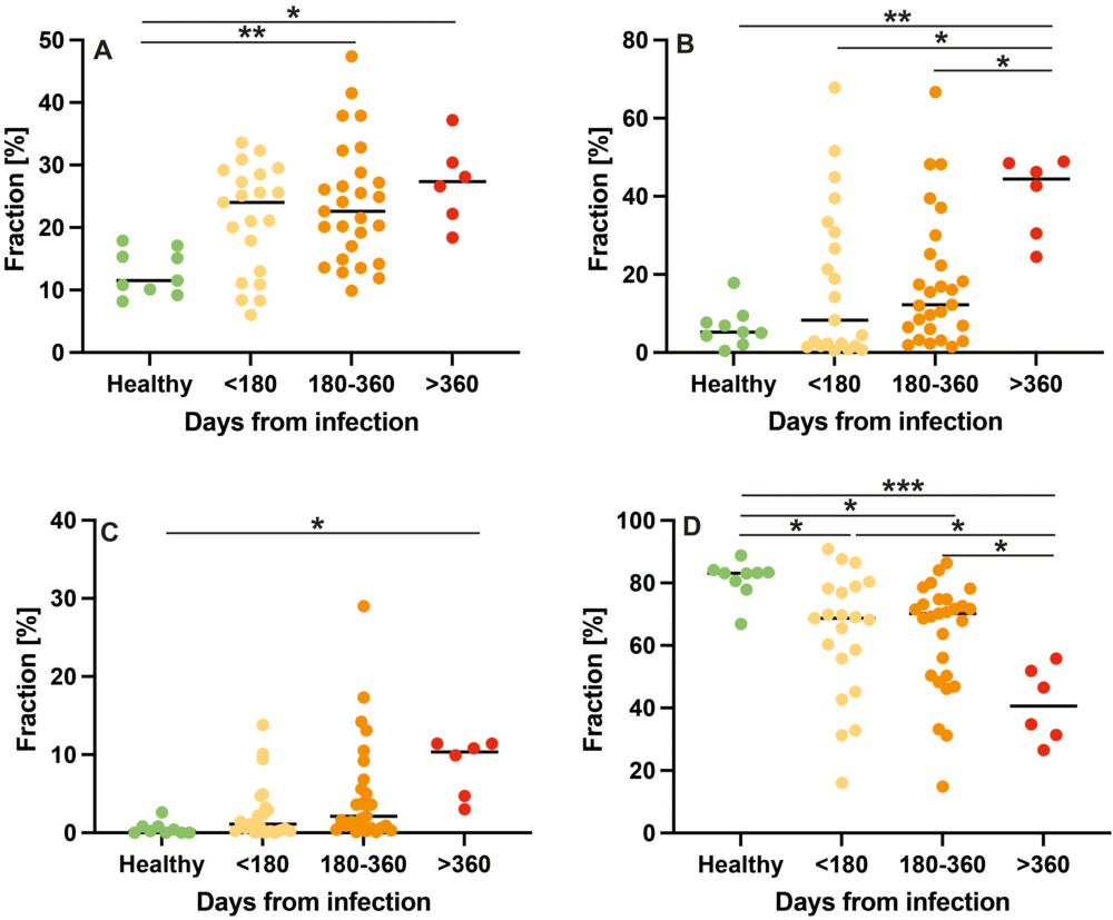
Figure 3. Comparison of low-field MRI parameters with respect to interval from first infection with dot plots for ventilation defects. The y-axis shows the fraction of defected or non-defected lung parenchyma on the automated measured axial plane. (A), perfusion defects (B), ventilation/perfusion match (no defects, C)) and ventilation/perfusion match (defect, D) in healthy controls, at less than 180 days, 180-360 days, and more than 360 days after SARS-CoV-2 infection.
High-res (TIF) version
(Right-click and Save As)