AI-based System Shows Promise in Tuberculosis Detection
Released: September 06, 2022
At A Glance
- Researchers have developed an AI deep-learning system that could quickly and automatically evaluate chest radiographs for TB.
- Researchers said the AI system may be able to aid screening in areas with limited resources.
- TB is an infectious disease of the lungs that kills more than a million people worldwide every year.
- RSNA Media Relations
1-630-590-7762
media@rsna.org - Linda Brooks
1-630-590-7738
lbrooks@rsna.org - Imani Harris
1-630-481-1009
iharris@rsna.org
OAK BROOK, Ill. — An artificial intelligence (AI) system detects tuberculosis (TB) in chest X-rays at a level comparable to radiologists, according to a study published in Radiology, a journal of the Radiological Society of North America (RSNA). Researchers said the AI system may be able to aid screening in areas with limited radiologist resources.
TB is an infectious disease of the lungs that kills more than a million people worldwide every year. The COVID-19 pandemic has exacerbated the problem, with recent reports indicating that 21% fewer people received care for TB in 2020 than in 2019. Almost 90% of the active TB infections occur in about 30 countries, many with scarce resources needed to address this public health problem.

“We have effective drugs for treating TB, but large-scale screening programs to detect TB are not always feasible in low-income countries due to cost and availability of expert radiologists,” said study co-author Rory Pilgrim, B.Eng., a product manager at Google Health AI in Mountain View, California.
Cost-effective TB screening using chest X-rays and AI has the potential to improve access to healthcare, Pilgrim said, particularly in difficult-to-reach populations.
“Bridging the expert shortage is where AI comes in,” said first author Sahar Kazemzadeh, B.S., software engineer at Google Health. “We can teach computers to recognize tuberculosis from X-rays so that in these low-resource settings a patient’s X-ray can be interpreted within seconds.”
Kazemzadeh and colleagues developed and assessed an AI system that can quickly and automatically evaluate chest X-rays for TB. The system uses deep learning, a type of AI that can be applied to teach the computer to recognize and predict medical conditions. The researchers developed the system using data from nine countries. They then tested it on data from five countries, covering multiple high-TB-burden countries, various clinical settings and a wide range of races and ethnicities. Over 165,000 images from more than 22,000 patients were used for model development and testing.

Analysis with 14 international radiologists showed that the deep-learning method was comparable to radiologists for the determination of active TB on chest X-rays.
“We wanted to see if this system predicts TB on par with radiologists, and that’s what the study is showing,” Pilgrim said. “AI performed really well with a variety of patients.”
Trends were similar across different patient subgroups, including a test set from gold miners in South Africa, a group with a high prevalence of TB, compared to the general public.
“What’s especially promising in this study is that we looked at a range of different datasets that reflected the breadth of TB presentation, different equipment and different clinical workflows,” Kazemzadeh said. “We found that this deep-learning system performs really well with all of them with a single operating point that was pre-selected based on a development dataset, something that other medical imaging AI systems have found challenging.”
If additional research supports the results, the deep-learning system could be used to automatically screen chest X-ray results for TB. People who test positive would then receive a sputum test or nucleic acid amplification testing (NAAT). These tests are relatively expensive, but if AI could filter the patients who need the test, the benefits would be extensive. Simulations using the deep-learning system to identify likely TB positive chest X-rays for NAAT confirmation reduced the cost by 40% to 80% per positive TB patient detected.
“By screening patients in the community and detecting TB before they get really sick, they could have better outcomes and may require a shorter course of treatment,” Pilgrim said. “Also, since TB is an infectious disease, if you can get to people early there will be less spread, compounding the benefits of this screening.”
The researchers are conducting work in Zambia in a prospective setting, meaning they are collecting data from patients attending screening, and providing NAAT for every patient for the purpose of studying the system. They also are looking at ways to get these models out to the world in a way that can have the maximum impact for patients.
“We hope this can be a tool used by non-expert physicians and healthcare workers to screen people en masse and get them to treatment where required without getting specialist doctors, who are in short supply,” Pilgrim said. “We believe we can do this with the people on the ground in a low-cost, high-volume way.”
“Deep Learning Detection of Active Pulmonary Tuberculosis at Chest Radiography Matched the Clinical Performance of Radiologists.” Collaborating with Pilgrim and Kazemzadeh were Jin Yu, M.S., Shahar Jamshy, M.Sc., Zaid Nabulsi, M.S., Christina Chen, M.D., Neeral Beladia, Ph.D., Charles Lau, M.D., Scott Mayer McKinney, M.S., Thad Hughes, Ph.D., Atilla P. Kiraly, Ph.D., Sreenivasa Raju Kalidindi, M.B.B.S., Monde Muyoyeta, Ph.D., Jameson Malemela, M.D., Ting Shih, M.S., Greg S. Corrado, Ph.D., Lily Peng, M.D., Ph.D., Katherine Chou, M.S., Po-Hsuan Cameron Chen, Ph.D., Yun Liu, Ph.D., Krish Eswaran, Ph.D., Daniel Tse, M.D., Shravya Shetty, M.S., and Shruthi Prabhakara, Ph.D.
Radiology is edited by David A. Bluemke, M.D., Ph.D., University of Wisconsin School of Medicine and Public Health, Madison, Wisconsin, and owned and published by the Radiological Society of North America, Inc. (https://pubs.rsna.org/journal/radiology)
RSNA is an association of radiologists, radiation oncologists, medical physicists and related scientists promoting excellence in patient care and health care delivery through education, research and technologic innovation. The Society is based in Oak Brook, Illinois. (RSNA.org)
For patient-friendly information on chest imaging, visit RadiologyInfo.org.
Images (JPG, TIF):
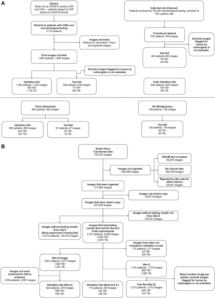
Figure 1. (A) Standards for Reporting of Diagnostic Accuracy Studies, or STARD, diagrams for validation and test data sets for Zambia, India, China, and the United States. (B) Standards for Reporting of Diagnostic Accuracy Studies diagram for validation and test data sets for South Africa. CAD = computer-aided detection, CIDRZ = Centre for Infectious Disease Research in Zambia, CXR = chest radiograph, GXP = GeneXpert (Cepheid), DICOM = Digital Imaging and Communications in Medicine, ID = identification, QA = quality assurance, TB = tuberculosis, TB– = TB negative, TB+ = TB positive. CAD4TB is a commercially available software (Delft Imaging).
High-res (TIF) version
(Right-click and Save As)
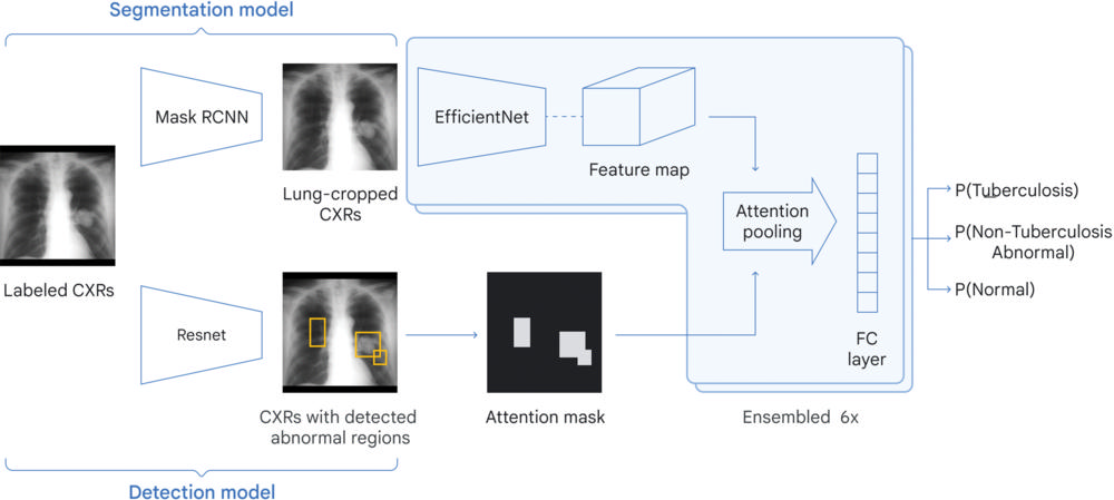
Figure 2. Overview of our deep learning system. The system consists of three modules: a lung cropping model to specifically crop the lungs, a detection model to identify regions of interest, and a classification model that takes the output from the other two models to predict the likelihood of the chest radiograph (CXR) being tuberculosis positive. The large-scale abnormality pretraining and noisy student semisupervised learning used to train the classification model are not shown here. “P” indicates probability, for example P(Tuberculosis) indicates the probability of the image showing signs of tuberculosis. FC = fully connected, RCNN = Region-based Convolutional Neural Network.
High-res (TIF) version
(Right-click and Save As)

Figure 3. Receiver operating characteristic (ROC) curves for the deep learning system (DLS) compared with radiologists on (A) a combined data set comprising four countries and (B) each data set individually. ROC curves for the DLS compared with radiologists on (C) subgroups based on HIV status in the Zambia data set and (D) an additional test data set from a mining population in South Africa. AUC = area under the ROC curve.
High-res (TIF) version
(Right-click and Save As)
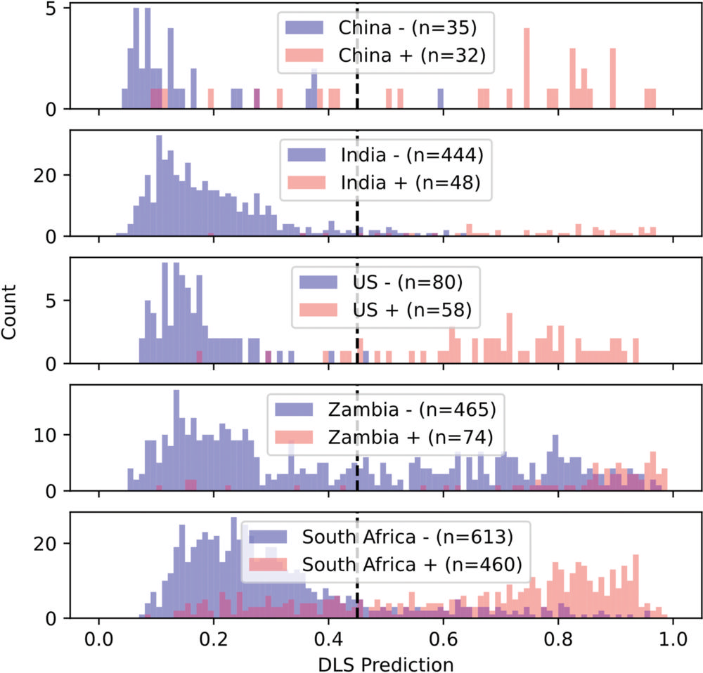
Figure 4. Histograms show the distribution of deep learning system (DLS) predictions stratified by positive (red) versus negative (blue) examples to illustrate shifts across data sets.
High-res (TIF) version
(Right-click and Save As)
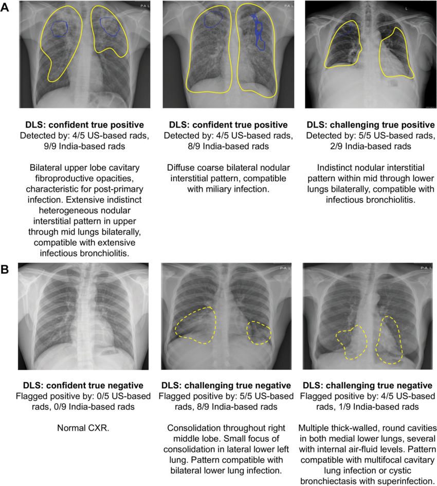
Figure 5. Examples of chest radiographs for which the deep learning system (DLS) provided the correct interpretation, corresponding to (A) tuberculosis (TB)–positive subjects and (B) TB-negative subjects. Blue outlines encircle salient regions via Grad-CAM (35) that most influence the positive prediction from the DLS and are shown when the DLS considered the image positive. Yellow outlines were annotated by a radiologist to indicate regions of interest; solid outlines indicate findings consistent with TB while dotted outlines indicate other findings. “Confident” indicates that the DLS predicted values close to 0 or 1, whereas “challenging” indicates that the DLS predicted values close to the operating point (0.45). CXR = chest radiograph, rads = radiologists
High-res (TIF) version
(Right-click and Save As)
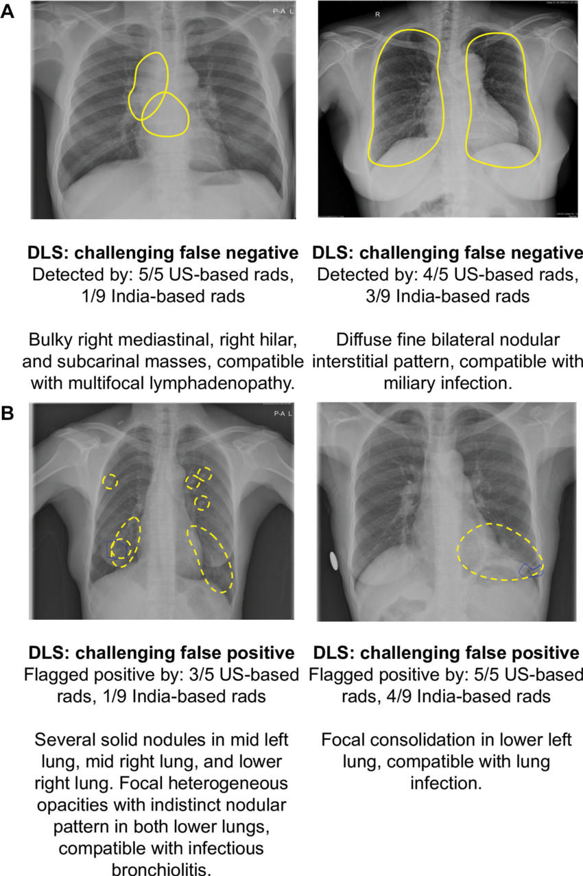
Figure 6. Examples of chest radiographs for which the deep learning system (DLS) provided the wrong interpretation, corresponding to (A) tuberculosis (TB)–positive subjects and (B) TB-negative subjects. Blue outlines encircle salient regions via Grad-CAM (35) that most influence the positive prediction from the DLS and are shown when the DLS considered the image positive. Yellow outlines were annotated by a radiologist to indicate regions of interest; solid outlines indicate findings consistent with TB while dotted outlines indicate other findings. “Confident” indicates that the DLS predicted values close to 0 or 1, whereas “challenging” indicates that the DLS predicted values close to the operating point (0.45). rads = Radiologists.
High-res (TIF) version
(Right-click and Save As)
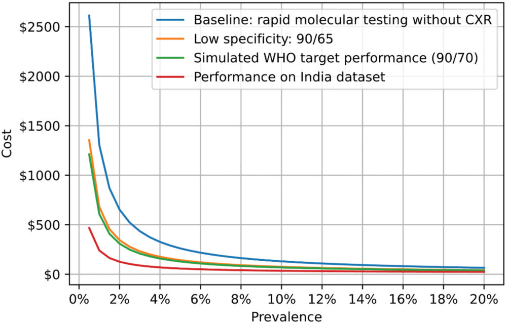
Figure 7. Graph shows the estimated cost per tuberculosis (TB)–positive patient detected using the deep learning system (DLS). Absolute cost on the y-axis represents the expected cost per TB-positive patient detected. For the other three cases, all cases underwent chest radiography and only the subset flagged as positive by a DLS with a certain sensitivity and specificity (described in the inset) underwent GeneXpert testing. The numbers 90/65 and 90/70 indicate the sensitivity and specificity; for example, 90/70 indicates 90% sensitivity and 70% specificity. WHO = World Health Organization.
High-res (TIF) version
(Right-click and Save As)