Emphysema Severity Associated with Higher Lung Cancer Risk
Released: May 03, 2022
At A Glance
- A new study links emphysema to a higher risk of lung cancer, a risk that increases with emphysema severity.
- Analysis of 21 studies involving more than 107,000 patients found a connection between CT assessments of emphysema and lung cancer.
- Emphysema is a chronic respiratory disease that has no cure, but many treatments are available to help manage symptoms.
- RSNA Media Relations
1-630-590-7762
media@rsna.org - Linda Brooks
1-630-590-7738
lbrooks@rsna.org - Imani Harris
1-630-481-1009
iharris@rsna.org
OAK BROOK, Ill. — CT-detected emphysema is linked to a higher risk of lung cancer, a risk that increases with emphysema severity, according to a new study published in the journal Radiology.

Lung cancer is the primary cause of cancer-related death worldwide, with more than 1 million deaths each year since 2000. However, lung cancer risk can be reduced by identifying treatable risk factors, such as chronic lung inflammation, together with smoking, genetics, diet, and occupational exposure.
Emphysema is a chronic respiratory disease characterized by damage to the alveoli, the tiny air sacs inside the lungs. Symptoms include shortness of breath, coughing with mucus, wheezing and chest tightness. There is no cure, but many treatments are available to help manage symptoms.
Emphysema shares many common risk factors with lung cancer, the leading cause of cancer-related deaths worldwide.
Cigarette smoking is one of the important shared risk factors of emphysema and lung cancer, as it enhances inflammation, DNA damage and accelerated aging. However, people with emphysema who’ve never smoked also have an increased risk of lung cancer, according to study co-author Marleen Vonder, Ph.D., from the Department of Epidemiology at University Medical Center Groningen in Groningen, the Netherlands.
“Other underlying mechanisms like genetic susceptibility, chronic inflammation or DNA damage and abnormal repair mechanisms, or a combination thereof, have been proposed to link emphysema and lung cancer,” she said.
For the new study, Dr. Vonder and colleagues identified studies from three large databases on the association between emphysema and lung cancer. Analysis of 21 studies involving more than 107,000 patients found a connection between visual and quantitative, or measurable, CT assessments of emphysema and lung cancer.
“Our meta-analysis showed that not only visually assessed but also quantitatively assessed emphysema on CT is associated with lung cancer and that this risk increases for more severe emphysema,” Dr. Vonder said.
While the findings support a link between the two devastating diseases, more research is needed before any changes are made to clinical care, Dr. Vonder said.
“It is too early to conclude whether the presence of CT-defined emphysema leads to incremental and independent prognostic value over that of already known shared risk factors of emphysema and lung cancer,” she said.
The associations between CT emphysema and lung cancer were higher for categories of visual assessment compared to quantitative assessment. Despite this finding, Dr. Vonder said that quantitative assessment may ultimately gain favor over visual assessment, as it can be fully automated. She and her colleagues are researching this approach and validating its use in specified populations.
“Potentially, emphysema detected on a baseline CT scan could be used to select high-risk participants who would require more frequent follow-up lung cancer screening,” Dr. Vonder said.
“Association between Chest CT-defined Emphysema and Lung Cancer: A Systematic Review and Meta-Analysis.” Collaborating with Dr. Vonder were Xiaofei Yang, M.D., Hendrik Joost Wisselink, M.Sc., Rozemarijn Vliegenthart, M.D., Ph.D., Marjolein A. Heuvelmans, M.D., Ph.D., Harry J. M. Groen, M.D., Ph.D., Monique D. Dorrius, M.D., Ph.D., and Geertruida H. de Bock, Ph.D.
Radiology is edited by David A. Bluemke, M.D., Ph.D., University of Wisconsin School of Medicine and Public Health, Madison, Wisconsin, and owned and published by the Radiological Society of North America, Inc. (https://pubs.rsna.org/journal/radiology)
RSNA is an association of radiologists, radiation oncologists, medical physicists and related scientists promoting excellence in patient care and health care delivery through education, research and technologic innovation. The Society is based in Oak Brook, Illinois. (RSNA.org)
For patient-friendly information on lung CT, visit RadiologyInfo.org.
Images (JPG, TIF):
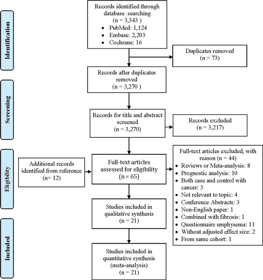
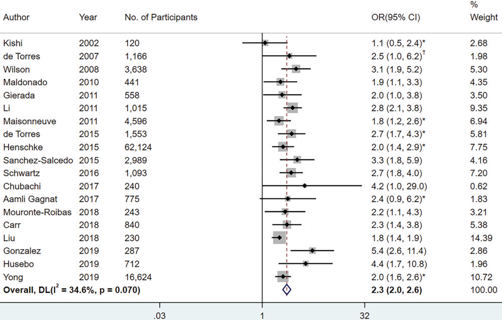
Figure 2. Forest plot of the random-effects meta-analysis for the association between emphysema (dichotomous variable) assessed visually and/or quantitatively with CT and lung cancer in 19 studies. The overall pooled odds ratio (OR) of emphysema for lung cancer was 2.3 (95% CI: 2.0, 2.6 [P < .001]). For the studies that assessed emphysema with two methods, only the ORs assessed with the main method were pooled in the overall estimates. Squares and horizontal lines represent estimates and 95% CIs, respectively, for each study part. Diamonds indicate pooled effect sizes with 95% CIs. DL = DerSimonian and Laird. * = Study reported hazard ratios. † = Study reported risk ratios.
High-res (TIF) version
(Right-click and Save As)
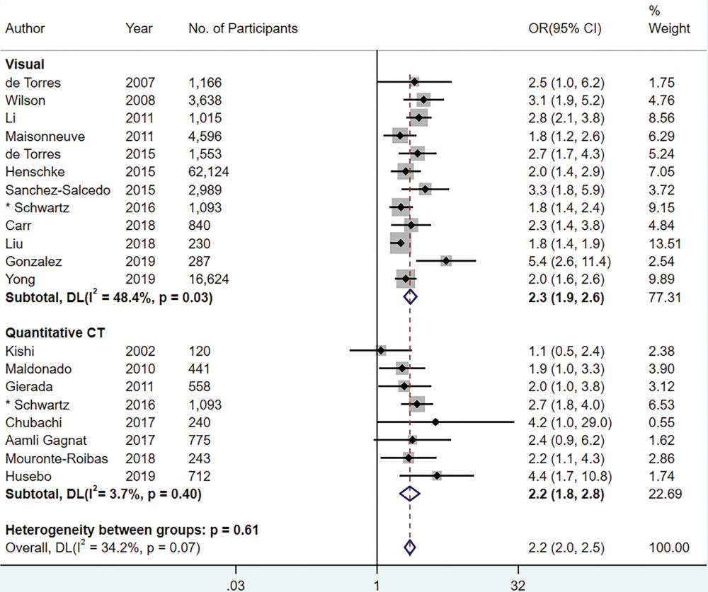
Figure 3. Forest plot of the random-effects meta-analysis for the association between emphysema and lung cancer, stratified by the emphysema assessment method. The pooled odds ratios (ORs) for lung cancer given visual and quantitative dichotomous emphysema assessment were 2.3 (95% CI: 1.9, 2.6 [P < .001]) and 2.2 (95% CI: 1.8, 2.8 [P < .001]), respectively. Squares and horizontal lines represent estimates and 95% CIs, respectively, for each study part. Diamonds indicate pooled effect sizes with 95% CIs. * = Study assessed emphysema both visually and quantitatively. DL = DerSimonian and Laird.
High-res (TIF) version
(Right-click and Save As)
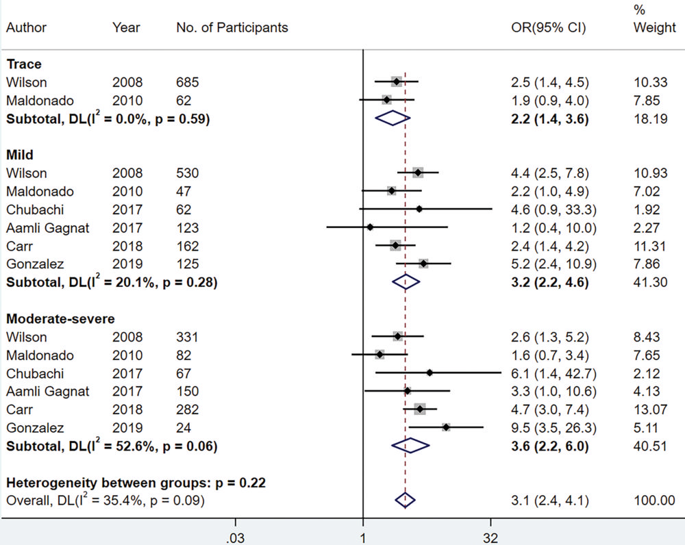
Figure 4. Forest plot of the random-effects meta-analysis for the association between emphysema severity (assessed visually and/or quantitatively) and lung cancer. The overall pooled odds ratios (ORs) of trace, mild, and moderate to severe emphysema for lung cancer were 2.2 (95% CI: 1.4, 3.6 [P = .001]), 3.2 (95% CI: 2.2, 4.6 [P < .001]) and 3.6 (95% CI: 2.2, 6.0 [P < .001]), respectively. Adjusted factors in these mixed-effects models varied, as shown in Table E2 (online). Squares and horizontal lines represent estimates and 95% CIs, respectively, for each study part. Diamonds indicate pooled effect sizes with 95% CIs. DL = DerSimonian and Laird.
High-res (TIF) version
(Right-click and Save As)
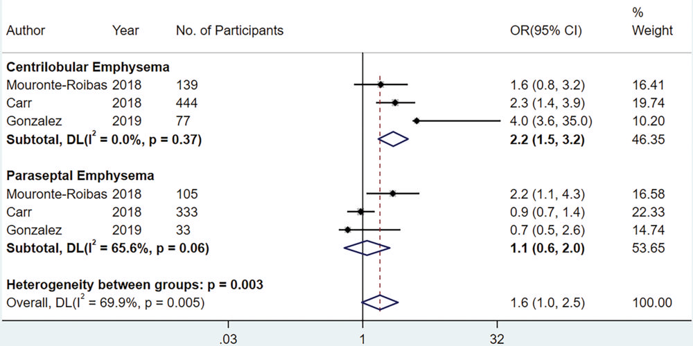
Figure 5. Forest plot of the random-effects meta-analysis for the association between emphysema subtype (assessed visually only) and lung cancer. The pooled odds ratios (ORs) for lung cancer odds in the presence of centrilobular and paraseptal emphysema were 2.2 (95% CI: 1.5, 3.2 [P < .001]) and 1.1 (95% CI: 0.6, 2.0 [P = .71]). Adjusted factors in these mixed-effects models varied, as shown in Table E2 (online). Squares and horizontal lines represent estimates and 95% CIs, respectively, for each study part. Diamonds indicate effect sizes with 95% CIs. DL = DerSimonian and Laird.
High-res (TIF) version
(Right-click and Save As)