COVID-19 Linked to Heart Inflammation in College Athletes
Released: November 29, 2021
At A Glance
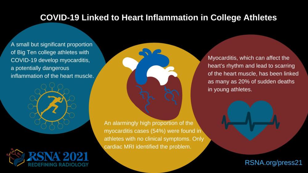
- A significant proportion of Big Ten college athletes with COVID-19 develop myocarditis, a potentially dangerous inflammation of the heart muscle.
- An alarmingly high proportion of the myocarditis cases (54%) were found in athletes with no clinical symptoms Only cardiac MRI identified the problem.
- Myocarditis, which can affect the heart’s rhythm and lead to scarring of the heart muscle, has been linked as many as 20% of sudden deaths in young athletes.
- RSNA Media Relations
1-630-590-7762
media@rsna.org - RSNA 2021 Newsroom
(Nov. 27-Dec. 2, 2021)
1-312-791-6610 - Linda Brooks
1-630-590-7738
lbrooks@rsna.org - Imani Harris
1-630-481-1009
iharris@rsna.org
CHICAGO — A small but significant percentage of college athletes with COVID-19 develop myocarditis, a potentially dangerous inflammation of the heart muscle, that can only be seen on cardiac MRI, according to a study being presented today at the annual meeting of the Radiological Society of North America (RSNA).
Myocarditis, which typically occurs as a result of a bacterial or viral infection, can affect the heart’s rhythm and ability to pump and often leaves behind lasting damage in the form of scarring to the heart muscle. It has been linked to as many as 20% of sudden deaths in young athletes. The COVID-19 pandemic raised concerns over an increased incidence of the condition in student-athletes.
For the new study, clinicians at schools in the highly competitive Big Ten athletic conference collaborated to collect data on the frequency of myocarditis in student-athletes recovering from COVID-19 infection. Conference officials had required all athletes who had COVID-19 to get a series of cardiac tests before returning to play, providing a unique opportunity for researchers to collect data on the athletes’ cardiac status.
Jean Jeudy, M.D., professor and radiologist at the University of Maryland School of Medicine in Baltimore, serves as the cardiac MRI core leader for the Big Ten Cardiac Registry. This registry oversaw the collection of all the data from the individual schools of the Big Ten conference.
Dr. Jeudy reviewed the results of 1,597 cardiac MRI exams collected at the 13 participating schools. There was no selection bias for cardiac MRI, as all COVID-positive athletes underwent a complete cardiac battery of tests including cardiac MRI, echocardiogram, ECG and blood tests, as well as a complete medical history.
Thirty-seven of the athletes, or 2.3%, were diagnosed with COVID-19 myocarditis, a percentage on par with the incidence of myocarditis in the general population. However, an alarmingly high proportion of the myocarditis cases were found in athletes with no clinical symptoms. Twenty of the patients with COVID-19 myocarditis (54%) had neither cardiac symptoms nor cardiac testing abnormalities. Only cardiac MRI identified the problem.
“Testing patients for clinical symptoms of myocarditis only captured a small percentage of all patients who had myocardial inflammation,” Dr. Jeudy said. “Cardiac MRI for all athletes yielded a 7.4-fold increase in detection.”
The implications of post-COVID-19 myocardial injury detected by cardiac MRI are still unknown.
“The main issue is the presence of persistent inflammation and/or myocardial scar,” Dr. Jeudy said. “Each of these can be an underlying foundation for additional damage and increased risk of arrhythmia.”
As part of the study, Dr. Jeudy and colleagues continue to add to the Big Ten Cardiac Registry to gain more understanding.
“We still don’t know the long-term effects,” Dr. Jeudy said. “Some athletes had issues that resolved within a month, but we also have athletes with continued abnormalities on their MRI as a result of their initial injury and scarring. There are a lot of chronic issues with COVID-19 that we need to know more about, and hopefully this registry can be one of the major parts of getting that information.”
The registry will allow researchers to look beyond the presence of abnormalities and study things like changes in exercise function over time.
“These are young patients, and the effects of myocardial inflammation can potentially impact their lives more significantly than in older patients,” Dr. Jeudy said. “That’s why we really want to push forward and continue to collect this data.”
Obstacles to widespread use of cardiac MRI in college athletes are significant and include cost and lack of access to advanced MRI capability at many centers. But, as the new study shows, cardiac MRI adds considerable value to cardiac testing.
“The role of cardiac MRI as a screening tool in this population needs to be explored,” Dr. Jeudy said. “The reality is that there are a small percentage of cases where we know the athletes have an increased risk for sudden death, and using cardiac MRI will increase the number of players who are identified.”
Note: Copies of RSNA 2021 news releases and electronic images will be available online at RSNA.org/press21.
RSNA is an association of radiologists, radiation oncologists, medical physicists and related scientists promoting excellence in patient care and health care delivery through education, research and technologic innovation. The Society is based in Oak Brook, Illinois. (RSNA.org)
Editor’s note: The data in these releases may differ from those in the published abstract and those actually presented at the meeting, as researchers continue to update their data right up until the meeting. To ensure you are using the most up-to-date information, please call the RSNA Newsroom at 1-312-791-6610.
For patient-friendly information on cardiac MRI, visit RadiologyInfo.org.
Images (JPG, TIF):

Figure 1. MRI shows late gadolinium enhancement (LGE) at the right ventricular attachment (arrow) of a 19-year-old male. Note: this image is for illustrative purposes only and is not associated with the Big Ten study group. Image courtesy of Radiology.
High-res (TIF) version
(Right-click and Save As)

Figure 2. LGE images, native T1 maps, extracellular volume maps, and global longitudinal strain (GLS) show myocardial abnormalities in adults recovering from moderate and severe COVID-19. Note: this image is for illustrative purposes only and is not associated with the Big Ten study group. Image courtesy of Radiology.
High-res (TIF) version
(Right-click and Save As)

Figure 3. MRI shows Prominent LGE involving more than 50% of the myocardium in the basal inferolateral and inferior segments of the left ventricle and in the anterolateral, inferolateral, and inferior segments of the left ventricle at the midventricular level in adolescent female. Note: this image is for illustrative purposes only and is not associated with the Big Ten study group. Image courtesy of Radiology: Cardiothoracic Imaging.
High-res (TIF) version
(Right-click and Save As)


