Imaging Speeds Lifesaving Care to Stroke Victims
Released: November 02, 2021
At A Glance
- A commonly available imaging technique can identify stroke patients most likely to benefit from a procedure to restore blood flow.
- A commonly available imaging technique can identify stroke patients most likely to benefit from a procedure to restore blood flow.
- Results showed a link between ischemic core growth and blood flow in the collateral blood vessels, with a symmetric collateral pattern strongly associated with slow growing, highly treatable ischemic cores.
- RSNA Media Relations
1-630-590-7762
media@rsna.org - Linda Brooks
1-630-590-7738
lbrooks@rsna.org
OAK BROOK, Ill. — A commonly available imaging technique can identify stroke patients most likely to benefit from a procedure to restore blood flow, a potential breakthrough in care that could save thousands of lives every year, according to a study in Radiology.
Large vessel occlusion strokes occur when a clot travels up from the heart or the neck and blocks blood flow in one of the large arteries providing blood to the brain. These strokes affect about 100,000 people in the United States every year and are responsible for most stroke-related deaths and long-term disability.
Endovascular thrombectomy, a treatment in which a tube is inserted into the blocked vessel to remove or destroy the clot, is highly effective for treating large vessel occlusions, but it has limitations.
“Thrombectomy is over 90% effective if we get to the patient in time,” said study co-first author R. Gilberto Gonzalez M.D., Ph.D., from the Department of Radiology at Massachusetts General Hospital (MGH) in Boston. “The problem is, we’re only treating less than 10% of the people who might benefit.”
One reason for the shortfall is the difficulty in the challenge in determining which patients need the treatment right away and which can wait—a function of how fast the brain injury, known as the ischemic core, is growing.
In a previous study, Dr. Gonzalez and colleagues discovered that the ischemic core grows very slowly in a significant subset of patients, meaning they can still benefit from thrombectomy 24 hours or more after the blockage occurs.
To learn more about how to identify these patients, Dr. Gonzalez, study co-first author Robert W. Regenhardt, M.D., Ph.D., a neuroendovascular fellow at MGH, and colleagues looked at results from 31 stroke patients who upon admission underwent computed tomography angiography (CTA), an imaging technique commonly available in hospitals. The patients also had MRIs at four time points over the next two days to track ischemic growth.
Results revealed a strong link between ischemic core growth and blood flow in the collateral blood vessels, smaller blood vessels recruited by the brain to make up for the loss of flow through the large vessels. A symmetric collateral pattern was strongly associated with slow growing, highly treatable ischemic cores.
“Our data shows that in almost half of patients, the core grows very slowly,” Dr. Gonzalez said. “That’s a huge number of people who are potentially treatable.”
Because CTA is widely available, patterns derived from it offer a potential lifeline for the tens of thousands of stroke victims who live in areas with no access to advanced imaging like CT perfusion and MRI. In theory, any person with a stroke who shows up at a hospital with limited facilities and gets CT and CTA could be quickly identified as having a good collateral circulation pattern, and then transferred to another hospital and treated.
“With CT angiography, we’ve found a way that’s widely available to identify these slow progressors,” Dr. Gonzalez said. “Now, a large number of patients with the most severe type of strokes can be treated.”
The researchers are currently working on an artificial intelligence algorithm that can automatically detect the specific patterns of collateral circulation associated with slower stroke progression. The algorithm can be run in the cloud so that any hospital can send data to it and get an evaluation within minutes.
“This method is very amenable to automation,” Dr. Gonzalez said. “This is how I see it having a major effect and benefitting tens of thousands of people every year.”
“Symmetric CTA Collaterals Identify Patients with Slow Progressing Stroke Likely to Benefit from Late Thrombectomy.” Collaborating with Drs. Gonzalez and Regenhardt were Julian He, M.D., Michael H. Lev, M.D., and Aneesh B. Singhal, M.D.
Radiology is edited by David A. Bluemke, M.D., Ph.D., University of Wisconsin School of Medicine and Public Health, Madison, Wisconsin, and owned and published by the Radiological Society of North America, Inc. (https://pubs.rsna.org/journal/radiology)
RSNA is an association of radiologists, radiation oncologists, medical physicists and related scientists promoting excellence in patient care and health care delivery through education, research and technologic innovation. The Society is based in Oak Brook, Illinois. (RSNA.org)
For patient-friendly information on CT and CTA, visit RadiologyInfo.org.
RSNA’s 107th Scientific Assembly and Annual Meeting, the world’s premier radiology forum, will be held at McCormick Place Chicago November 28 – December 2. #RSNA21
Images (JPG, TIF):
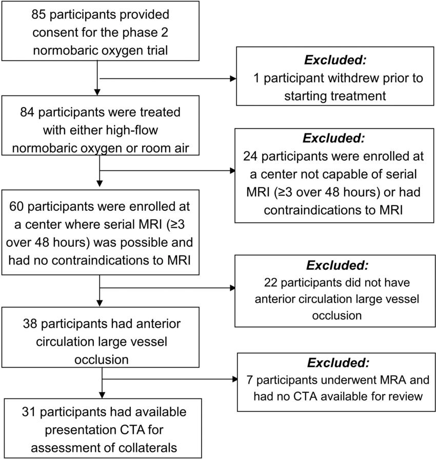
Figure 1. Flowchart shows reasons patients were excluded from this study. CTA = CT angiography, MRA = MR angiography.
High-res (TIF) version
(Right-click and Save As)
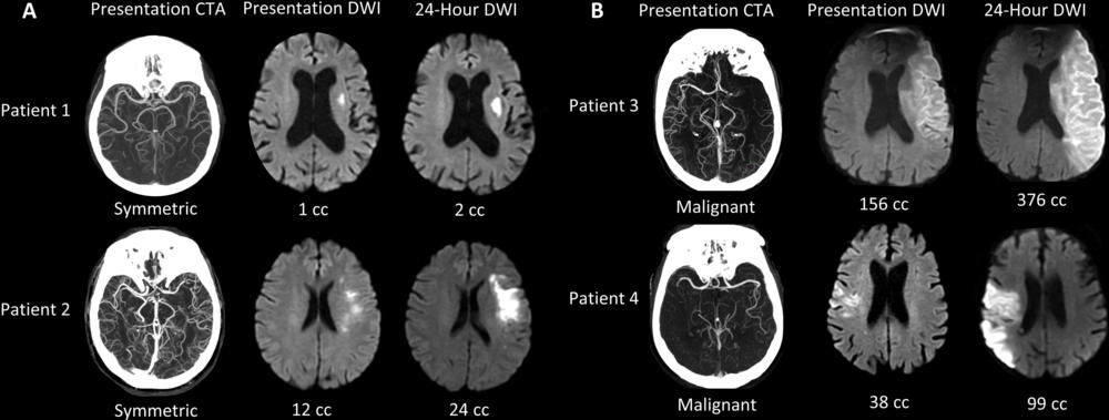
Figure 2. Collateral patterns at presentation CT angiography (CTA) and corresponding ischemic cores at presentation and 24-hour diffusion-weighted imaging (DWI). (A) Examples of patients with symmetric collaterals. (B) Examples of patients with malignant collaterals. Patient 1 is an 84-year-old woman with National Institutes of Health Stroke Scale (NIHSS) score of 7 and symptom onset 4.1 hours prior. Patient 2 is a 70-year-old woman with NIHSS score of 7 and symptom onset 5.6 hours prior. Patient 3 is a 71-year-old man with NIHSS score of 21 and symptom onset 10.8 hours prior. Patient 4 is an 85-year-old man with NIHSS score of 6 and symptom onset 2.5 hours prior.
High-res (TIF) version
(Right-click and Save As)
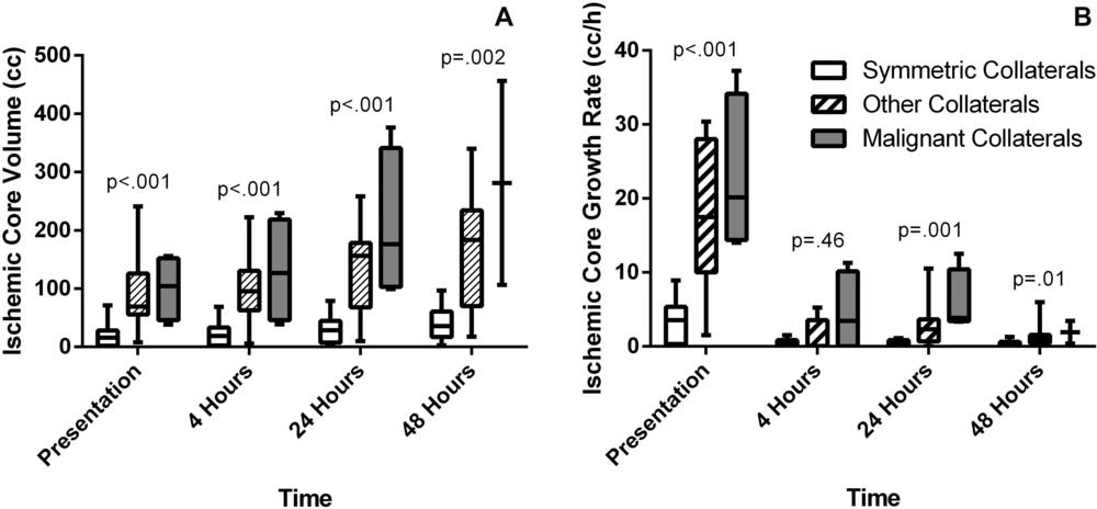
Figure 3. Collateral patterns at presentation CT angiography (CTA) have different ischemic core volumes and ischemic core growth rates at multiple time points. (A) Box-and-whisker plots with Kruskal Wallis test show that at presentation, median ischemic core volume was 16 cm3 for symmetric collaterals, 69 cm3 for other collaterals, and 104 cm3 for malignant collaterals. At 4 hours, median ischemic core volume was 19 cm3 for symmetric collaterals, 95 cm3 for other collaterals, and 127 cm3 for malignant collaterals. At 24 hours, median ischemic core volume was 28 cm3 for symmetric collaterals, 156 cm3 for other collaterals, and 176 cm3 for malignant collaterals. At 48 hours, median ischemic core volume was 36 cm3 for symmetric collaterals, 183 cm3 for other collaterals, and 281 cm3 for malignant collaterals. (B) Box-and-whisker plots with Kruskal Wallis test show that at presentation, median ischemic core growth rate (IGR) was 4 cm3 per hour for symmetric collaterals, 17 cm3 per hour for other collaterals, and 20 cm3 per hour for malignant collaterals. At 4 hours, median IGR was 0 cm3 per hour for symmetric collaterals, 0 cm3 per hour for other collaterals, and 3 cm3 per hour for malignant collaterals. At 24 hours, median IGR was 1 cm3 per hour for symmetric collaterals, 2 cm3 per hour for other collaterals, and 4 cm3 per hour for malignant collaterals. At 48 hours, median IGR was 0 cm3 per hour for symmetric collaterals, 1 cm3 per hour for other collaterals, and 2 cm3 per hour for malignant collaterals.
High-res (TIF) version
(Right-click and Save As)
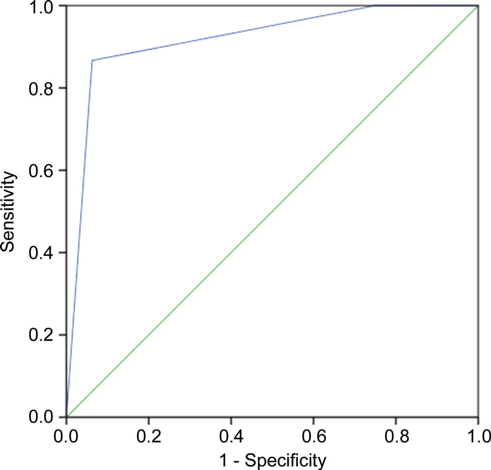
Figure 4. Collateral pattern can predict 24-hour ischemic core volume less than 50 cm3. Receiver operator characteristic curve (blue line; area under the receiver operating characteristic curve, 0.92; 95% CI: 0.81, 1.00; P < .001) showed symmetric collaterals had a sensitivity of 87% (13 of 15) and a specificity of 94% (15 of 16); nonmalignant collaterals had a sensitivity of 100% (15 of 15) and a specificity of 25% (four of 16). Green line indicates the line of no-discrimination.
High-res (TIF) version
(Right-click and Save As)