Digital Breast Tomosynthesis Reduces Rate of Interval Cancers
Released: April 06, 2021
At A Glance
- DBT reduces the rate of interval breast cancers (cancers that are diagnosed between screenings) compared to screening with only digital mammography.
- Interval cancer rates with DBT and digital mammography was 1.6 per 1,000 screened, compared to 2.8 per 1000 with digital mammography only.
- Interval cancers are generally more aggressive than cancers detected during a screening exam.
- RSNA Media Relations
1-630-590-7762
media@rsna.org - Linda Brooks
1-630-590-7738
lbrooks@rsna.org - Katherine Anderson
1-630-491-1009
kanderson@rsna.org
OAK BROOK, Ill. — Screening with digital breast tomosynthesis (DBT) reduces the rate of interval breast cancers compared to screening with digital mammography, according to a study published in Radiology. The study adds to a growing body of evidence supporting DBT as a breast cancer screening tool with important advantages over mammography.

DBT works by capturing a series of X-ray images of the breast from different angles. Previous research has shown that it has a higher sensitivity for breast cancer detection than digital mammography.
The impact of these additional DBT-detected cancers is not fully understood. While they may constitute a screening benefit, they could also contribute to overdiagnosis, a term for the diagnosis of early-stage, slow-growing cancers that would not have caused harm to the patient in their lifetime.
The rate of interval cancers—cancers that arise between routine screenings—offers one way to better elucidate screening benefits. They are considered more aggressive than cancers detected during a screening exam.
“Interval cancers have, in general, a more aggressive biological profile than screen-detected cancers,” said study lead author Kristin Johnson, M.D., radiology resident at Skåne University Hospital in Malmö, and Ph.D. student at Lund University, Sweden. “This means that the prognosis is less favorable for interval cancers compared to screen-detected cancers.”
Interval cancer detection rate reporting is required in many screening programs as an indicator of effectiveness. A reduction in the interval cancer rate when using DBT might be attributed to improved detection of rapidly growing cancers with poorer prognosis, possibly contributing to lower breast cancer mortality.
For the new study, Dr. Johnson and colleagues compared interval cancer rates in Sweden’s population-based Malmö Breast Tomosynthesis Screening Trial with those from an age-matched control group of patients who underwent digital mammography at the same center.
The study group included almost 15,000 women who were screened with DBT and digital mammography between 2010 and 2015. Those women were matched with a control group of more than 26,000 women who had only digital mammography screening during the same time period.
The interval cancer rate in the patients screened with DBT and digital mammography was 1.6 per 1,000 screened, significantly lower than 2.8 per 1000 in the group screened with digital mammography only. The interval cancers in the trial generally had non-favorable characteristics.
The reduced interval cancer rate after screening with DBT could translate into screening benefits, according to Dr. Johnson.
“One could speculate that some of the additional cancers detected in DBT screening would have been diagnosed as interval cancers if not detected by DBT,” she said.
The results support the growing evidence of DBT as a screening modality with potential to replace digital mammography in future breast cancer screening. However, Dr. Johnson cautioned that other trials have not shown significantly reduced interval cancer rates in DBT screening compared to digital mammography screening. And interval cancer rates, while important, are not the only measure when evaluating the potential benefits from DBT in screening.
“Other factors, such as cancer types detected and cost-benefit, have to be taken into account,” Dr. Johnson said.
Toward that end, the researchers are working on a cost-benefit analysis of the Malmö Breast Tomosynthesis Screening Trial. They are also analyzing the trial for false positive recalls, those instances when patients are called back for additional screening for suspicious findings that end up being benign.
“Interval Breast Cancer Rates and Tumor Characteristics in the Prospective Population-based Malmö Breast Tomosynthesis Screening Trial.” Collaborating with Dr. Johnson were Kristina Lång, M.D., Ph.D., Debra M. Ikeda, M.D., Anna Åkesson, B.S., Ingvar Andersson, M.D., Ph.D., and Sophia Zackrisson, M.D., Ph.D.
Radiology is edited by David A. Bluemke, M.D., Ph.D., University of Wisconsin School of Medicine and Public Health, Madison, Wisconsin, and owned and published by the Radiological Society of North America, Inc. (https://pubs.rsna.org/journal/radiology)
RSNA is an association of radiologists, radiation oncologists, medical physicists and related scientists promoting excellence in patient care and health care delivery through education, research and technologic innovation. The Society is based in Oak Brook, Illinois. (RSNA.org)
For patient-friendly information on breast imaging, visit RadiologyInfo.org.
Images (JPG, TIF):
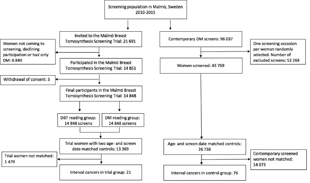
Figure 1. Flowchart of population and design in the Malmö Breast Tomosynthesis Screening Trial and the contemporary population. DBT = digital breast tomosynthesis, DM = digital mammography.
High-res (TIF) version
(Right-click and Save As)
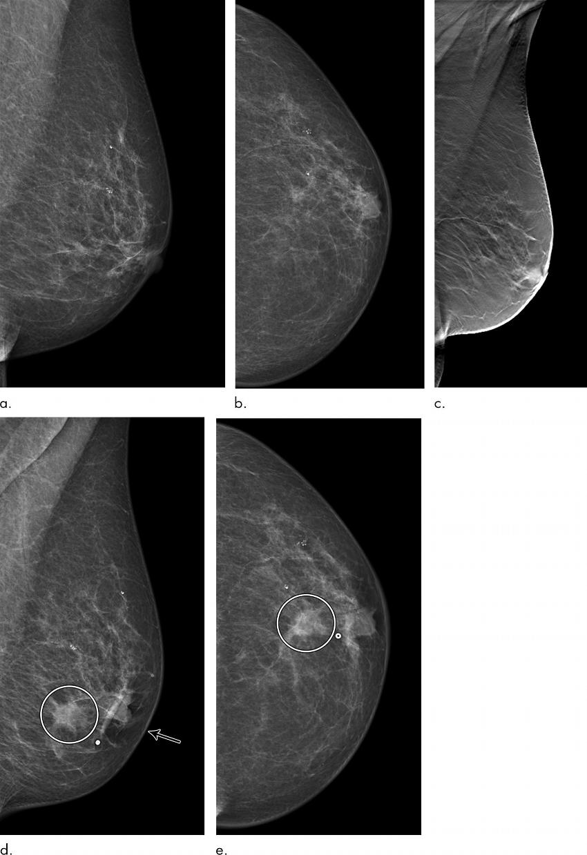
Figure 2. Images in a 72-year-old woman who was diagnosed with a 13-mm lymph node-negative invasive lobular carcinoma luminal B–like human epidermal growth factor receptor 2 breast cancer 18 months after a screening negative for cancer in the Malmö Breast Tomosynthesis Screening Trial. (a) Mediolateral oblique and (b) craniocaudal digital mammography (DM) images at screening. The slight retraction of the nipple was unchanged compared with previous DM screening images. (c) Digital breast tomosynthesis at screening. DM images of (d) mediolateral oblique and (e) craniocaudal views at diagnosis, small marker at lump location. Increased nipple retraction (arrow) and central mass (circle on d and e).
High-res (TIF) version
(Right-click and Save As)
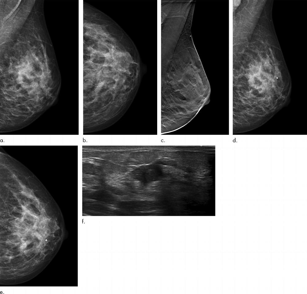
Figure 3. Images in a 43-year-old woman diagnosed with a 13-mm lymph node–positive invasive ductal carcinoma luminal B–like human epidermal growth factor receptor 2 breast cancer 7 months after a screening negative for cancer in the Malmö Breast Tomosynthesis Screening Trial. (a) Mediolateral and (b) craniocaudal digital mammography (DM) images at screening. (c) Digital breast tomosynthesis screening image. (d) Mediolateral and (e) craniocaudal DM images at diagnosis with a small marker at lump location. The breast cancer is difficult to distinguish. (f) US image at diagnosis; breast cancer clearly visible.
High-res (TIF) version
(Right-click and Save As)