Novel Technique ‘Stuns’ Arthritis Pain in Shoulder and Hip
Released: November 16, 2020
At A Glance
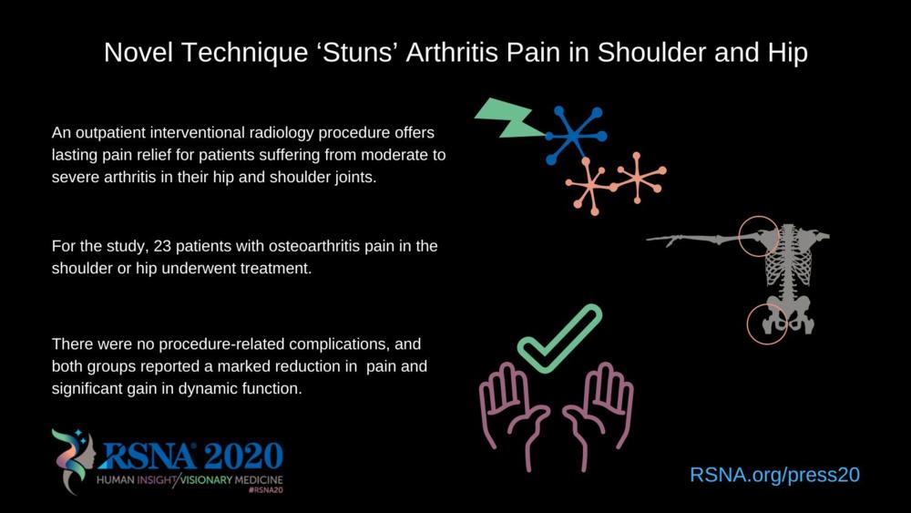
- An outpatient interventional radiology procedure offers lasting pain relief for patients suffering from moderate to severe arthritis in their hip and shoulder joints.
- For the study, 23 patients with osteoarthritis pain in the shoulder or hip underwent treatment.
- There were no procedure-related complications, and both groups reported a marked reduction in pain and significant gain in dynamic function.
- RSNA Media Relations
1-630-590-7762
media@rsna.org - Linda Brooks
1-630-590-7738
lbrooks@rsna.org
OAK BROOK, Ill. — A novel outpatient procedure offers lasting pain relief for patients suffering from moderate to severe arthritis in their hip and shoulder joints, according to a study presented at the annual meeting of the Radiological Society of North America (RSNA). Researchers said the procedure could help reduce reliance on addictive opiates.
People with moderate to severe pain related to osteoarthritis face limited treatment options. Common approaches like injections of anesthetic and corticosteroids into the affected joints grow less effective as the arthritis progresses and worsens.
“Usually, over time patients become less responsive to these injections,” said Felix M. Gonzalez, M.D., from the Radiology Department at Emory University School of Medicine in Atlanta, Georgia. “The first anesthetic-corticosteroid injection may provide six months of pain relief, the second may last three months, and the third may last only a month. Gradually, the degree of pain relief becomes nonsignificant.”
Without pain relief, patients face the possibility of joint replacement surgery. Many patients are ineligible for surgery because of health reasons, whereas many others choose not to go through such a major operation. For those patients, the only other viable option may be opiate painkillers, which carry the risk of addiction.
Dr. Gonzalez and colleagues have been studying the application of a novel interventional radiology treatment known as cooled radiofrequency ablation (c-RFA) to achieve pain relief in the setting of advanced degenerative arthritis. The procedure involves the placement of needles where the main sensory nerves exist around the shoulder and hip joints. The nerves are then treated with a low-grade current known as radiofrequency that “stuns” them, slowing the transmission of pain to the brain.
For the new study, 23 people with osteoarthritis underwent treatment, including 12 with shoulder pain and 11 with hip pain that had become unresponsive to anti-inflammatory pain control and intra-articular lidocaine-steroid injections. Treatment was performed two to three weeks after the patients received diagnostic anesthetic nerve blocks. The patients then completed surveys to measure their function, range of motion and degree of pain before and at three months after the ablation procedures.
There were no procedure-related complications, and both the hip and shoulder pain groups reported statistically significant decrease in the degree of pain with corresponding increase in dynamic function after the treatment.
“In our study, the results were very impressive and promising,” Dr. Gonzalez said. “The patients with shoulder pain had a decrease in pain of 85%, and an increase in function of approximately 74%. In patients with hip pain, there was a 70% reduction in pain, and a gain in function of approximately 66%.”
The procedure offers a new alternative for patients who are facing the prospect of surgery. In addition, it can decrease the risk of opiate addiction.
“This procedure is a last resort for patients who are unable to be physically active and may develop a narcotic addiction,” Dr. Gonzalez said. “Until recently, there was no other alternative for the treatment of patients at the end of the arthritis pathway who do not qualify for surgery or are unwilling to undergo a surgical procedure.”
At last year’s RSNA annual meeting, Dr. Gonzalez presented similarly encouraging results from a study of a similar procedure for the treatment of knee arthritis. Together, the knee, shoulder and hip articulations account for approximately 95% of all arthritis cases.
The procedure could have numerous applications outside of treating arthritic pain, Dr. Gonzalez explained. Potential uses include treating pain related to diseases like cancer and sickle cell anemia-related pain syndrome, for example.
“We’re just scratching the surface here,” Dr. Gonzalez said. “We would like to explore efficacy of the treatment on patients in other settings like trauma, amputations and especially in cancer patients with metastatic disease.”
Co-authors are Omar N. Kallas, M.D., Nima Kokabi, M.D., Zachary Bercu, M.D., David Reiter, Monica B. Umpierrez, M.D., Yi N. Guo, Adam D. Singer, M.D., Janice M. Newsome, M.D., Shervin Oskouei, Nickolas Reimer, and Philip K. Wong, M.D.
For more information and images, visit RSNA.org/press20. Press account required to view embargoed materials.
RSNA is an association of radiologists, radiation oncologists, medical physicists and related scientists promoting excellence in patient care and health care delivery through education, research and technologic innovation. The Society is based in Oak Brook, Illinois. (RSNA.org)
Editor’s note: The data in these releases may differ from those in the published abstract and those actually presented at the meeting, as researchers continue to update their data right up until the meeting. To ensure you are using the most up-to-date information, please call the RSNA media relations team at Newsroom at 1-630-590-7762.
For patient-friendly information on radiofrequency ablation, visit RadiologyInfo.org.
Video (MP4):

Video 1. Dr. Felix M. Gonzalez discusses his research on cooled radiofrequency ablation for the treatment of shoulder and hip osteoarthritis.
Download MP4
(Right-click and Save As)
Video 2. Animation of cooled radiofrequency ablation procedure to treat hip osteoarthritis.
Download MP4
(Right-click and Save As)
Images (JPG, TIF):

Figure 1. Differences between cooled radiofrequency ablation and conventional standard radiofrequency ablation.
High-res (TIF) version
(Right-click and Save As)

Figure 2. Cooled radiofrequency ablation components.
High-res (TIF) version
(Right-click and Save As)

Figure 3. Shoulder-specific technique: there are five ablation targets
High-res (TIF) version
(Right-click and Save As)

Figure 4. Suprascapular nerve cooled radiofrequency ablation targets.
High-res (TIF) version
(Right-click and Save As)

Figure 5. Suprascapular nerve cooled radiofrequency ablation: Target #1
High-res (TIF) version
(Right-click and Save As)

Figure 6. Suprascapular nerve cooled radiofrequency ablation: Target #2
High-res (TIF) version
(Right-click and Save As)
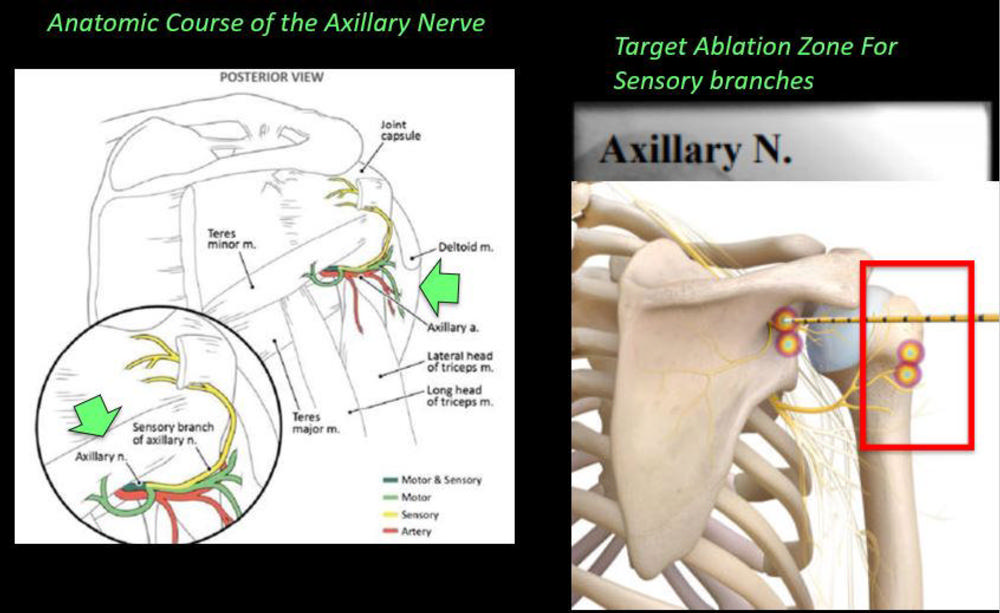
Figure 7. Axillary nerve cooled radiofrequency ablation: ablation targets.
High-res (TIF) version
(Right-click and Save As)

Figure 8. Axillary nerve cooled radiofrequency ablation: Ablation target #3.
High-res (TIF) version
(Right-click and Save As)
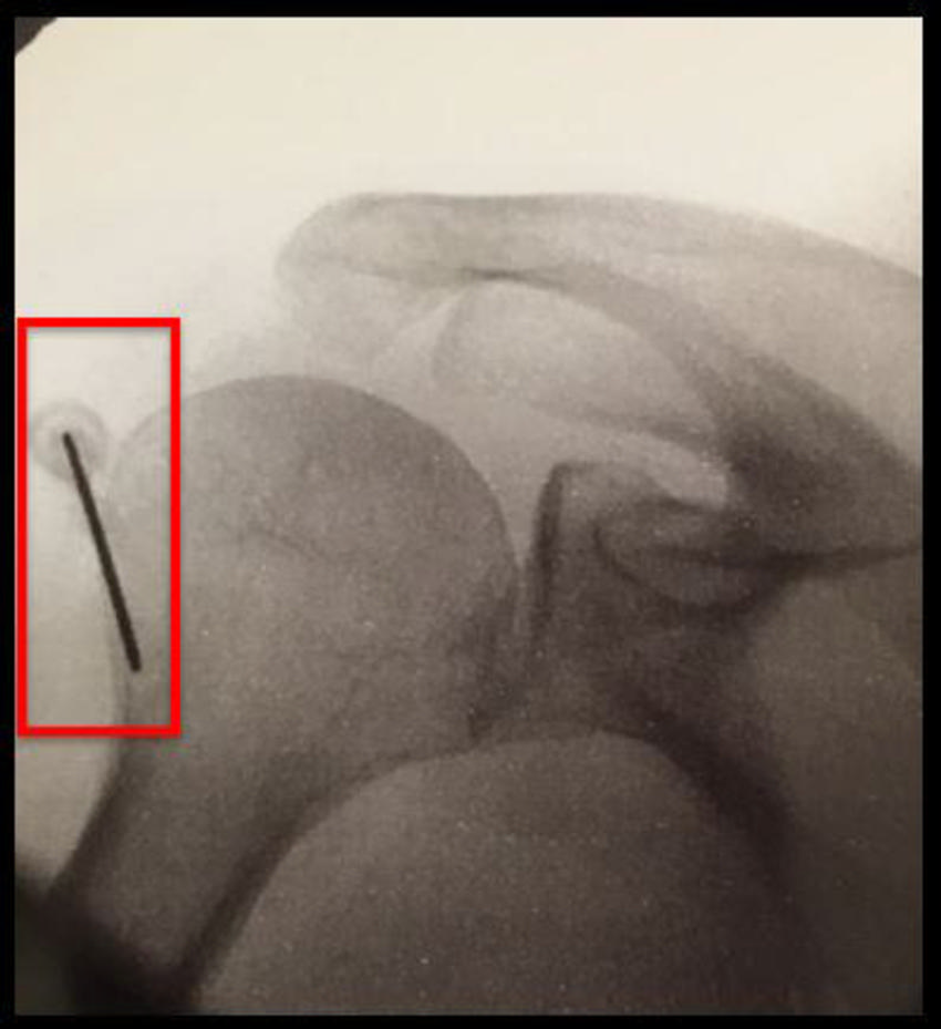
Figure 9. Axillary nerve cooled radiofrequency ablation: Ablation target #4.
High-res (TIF) version
(Right-click and Save As)
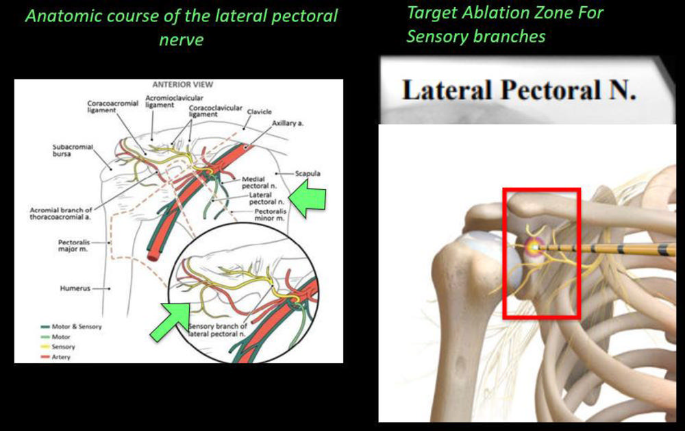
Figure 10. Lateral pectoral nerve cooled radiofrequency ablation: Ablation target.
High-res (TIF) version
(Right-click and Save As)
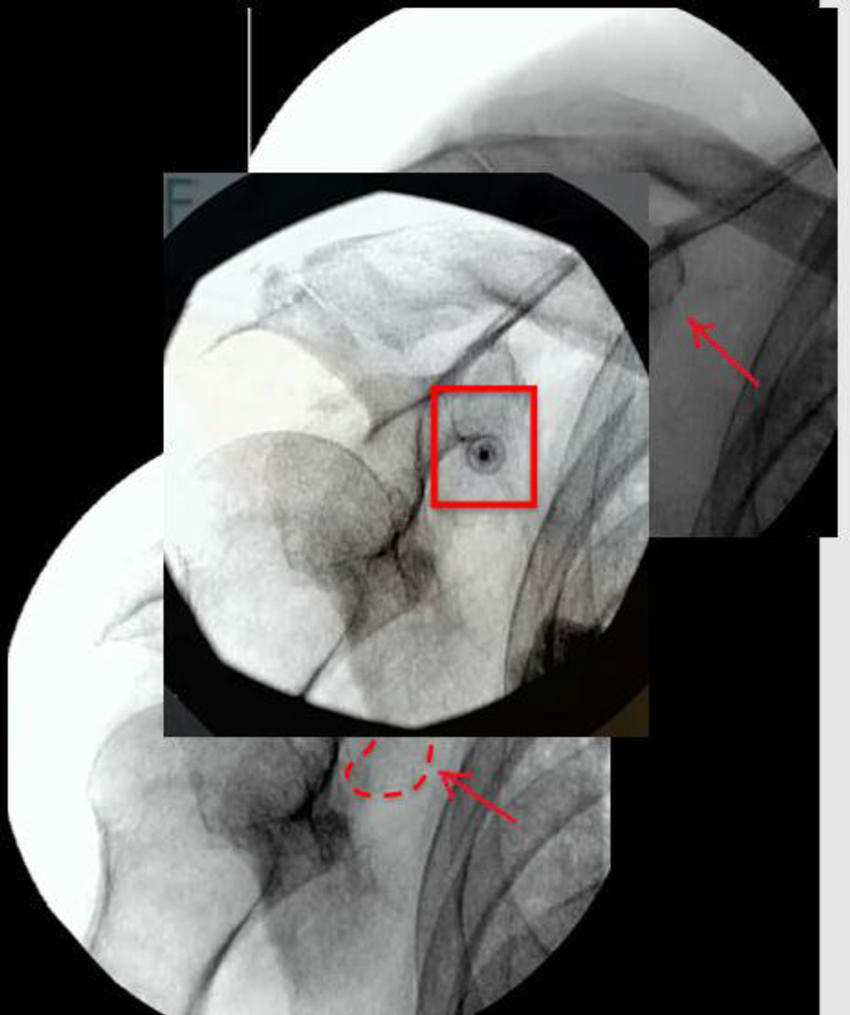
Figure 11. Lateral pectoral nerve: Ablation target #5
High-res (TIF) version
(Right-click and Save As)
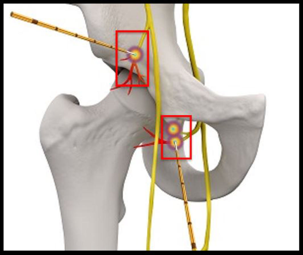
Figure 12. Hip-specific cooled radiofrequency ablation technique: There are three ablation targets.
High-res (TIF) version
(Right-click and Save As)
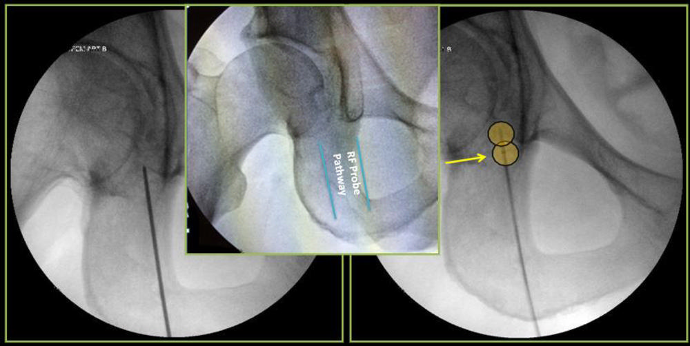
Figure 13. Hip cooled radiofrequency ablation: Obturator nerve ablation.
High-res (TIF) version
(Right-click and Save As)
Powerpoint:


