Vaping Impairs Vascular Function
Released: August 20, 2019
At A Glance
- Vaping immediately impacts vascular function, even when the solution does not include nicotine.
- Researchers performed MRI exams on 31 healthy non-smoking adults before and after nicotine-free e-cigarette inhalation.
- In 2018 more than 3.6 million middle and high school students reported using e-cigarettes.
- RSNA Media Relations
1-630-590-7762
media@rsna.org - Linda Brooks
1-630-590-7738
lbrooks@rsna.org - Dionna Arnold
1-630-590-7791
darnold@rsna.org
OAK BROOK, Ill. — Inhaling a vaporized liquid solution through an e-cigarette, otherwise known as vaping, immediately impacts vascular function even when the solution does not include nicotine, according to the results of a new study published in Radiology.
E-cigarette use is on the rise. According to the Centers for Disease Control and Prevention, more than 9 million adults in the U.S. use e-cigarettes, and vaping has become especially popular among teens. The 2018 National Youth Tobacco Survey reported that in 2018 more than 3.6 million middle and high school students were using e-cigarettes.
"The use of e-cigarettes is a current public health issue because of widespread use, especially among teenagers, and the fact that the devices are advertised as safe despite uncertainty about the effects of long-term use," said Alessandra Caporale, Ph.D., a post-doctoral researcher in the Laboratory for Structural, Physiologic and Functional Imaging (LSPFI) directed by senior author and principal investigator of the study, Felix W. Wehrli, Ph.D., at the University of Pennsylvania Perelman School of Medicine in Philadelphia. The research was funded by the National Heart, Lung, and Blood Institute (NHLBI).
According to the authors, e-cigarette inhalants, upon vaporization of the e-cigarette solution, contain potentially harmful toxic substances. Once inhaled, these particles can reach the alveoli of the lung, from where they are taken up by the blood vessels, thereby interfering with vascular function and promoting inflammation.
To study the acute effects of vaping on systemic vascular function, the researchers performed a series of MRI exams on 31 healthy non-smoking young adults (mean age 24; 14 women) before and after nicotine-free e-cigarette inhalation. The e-cigarette liquid contained pharma-grade propylene glycol and glycerol with flavoring, but no nicotine.
Using novel multi-parametric MRI protocols developed by Michael C. Langham, Ph.D., one of the co-authors of the study, scans of the femoral artery in the leg, the aorta and brain were performed before and after a single vaping episode equivalent to smoking a single conventional cigarette. For the femoral artery MRI, blood flow in the upper leg was constricted using a cuff and then released; the brain MRI was conducted in the sagittal sinus, during a series of thirty-second breath holds and normal breathing.
Comparing the pre- and post-MRI data, the single episode of vaping resulted in reduced blood flow and impaired vascular reactivity in the femoral artery, in which a 34 percent reduction in flow-mediated dilation—or the dilation of an artery mediated by blood flow increase—was found. There was a 17.5 percent reduction in peak flow, a 25.8 percent reduction in blood acceleration.
These findings suggest impaired function of the endothelium (inner lining of blood vessels). Moreover, a 20 percent reduction in venous oxygen saturation is indicative of altered microvascular function. The researchers also found a three percent increase in aortic pulse-wave velocity, a measure of arterial stiffness, or the rate at which pressure waves move down the aorta.
"These products are advertised as not harmful, and many e-cigarette users are convinced that they are just inhaling water vapor," Dr. Caporale said. "But the solvents, flavorings and additives in the liquid base, after vaporization, expose users to multiple insults to the respiratory tract and blood vessels."
Dr. Caporale said further studies are needed to address the potentially adverse long-term effects of vaping on vascular health.
"Acute Effects of Electronic Cigarette Aerosol Inhalation on Vascular Function Detected at Quantitative MRI." Collaborating with Drs. Caporale, Wehrli and Langham were Wensheng Guo, Ph.D., Alyssa Johncola, B.A., and Shampa Chatterjee, Ph.D.
Radiology is edited by David A. Bluemke, M.D., Ph.D., University of Wisconsin School of Medicine and Public Health, Madison, Wis., and owned and published by the Radiological Society of North America, Inc. (http://radiology.rsna.org/)
RSNA is an association of over 53,400 radiologists, radiation oncologists, medical physicists and related scientists, promoting excellence in patient care and health care delivery through education, research and technologic innovation. The Society is based in Oak Brook, Ill. (RSNA.org)
For patient-friendly information on MRI, visit RadiologyInfo.org.
Images (.JPG and .TIF format)

Figure 1. Flowchart of participant enrollment and exclusion, completed assessments, and test-retest repeatability. aPWV = aortic pulse wave velocity, BMI = body mass index, CVR = cerebrovascular reactivity, PVR = peripheral vascular reactivity.
High-res (TIF) version
(Right-click and Save As)

Figure 2. Response to cuff occlusion in the femoral circulation. A, Vessel-wall images of the superficial femoral artery (SFA) at different points (60 seconds [A60], 90 seconds [A90], and 120 seconds [A120]) as indicated by crosses in B during reactive hyperemia. The dashed circles represent the lumen area at baseline. B, Superficial femoral vein (SFV) oxygen saturation (SvO2) at baseline (green line) and during hyperemia. C, SFA blood flow velocity (V). D, Axial image obtained with MRI in the thigh, with SFA and SFV indicated in red and blue, respectively. Sample data shown for a representative participant. ΔSvO2 = peak-to-peak SvO2, HI = hyperemic index, PFR = peripheral flow reserve, RI = resistivity index, TFF = time of forward flow, TP = time to peak, TW = washout time, Vb = baseline velocity, VP = peak hyperemic velocity, Vr = retrograde velocity during early diastole, Vs = systolic velocity.
High-res (TIF) version
(Right-click and Save As)

Figure 3. Neurovascular response to breath hold. (a) Magnitude image intensity of superior sagittal sinus (SSS, box). Insets show velocity maps at different points of the velocity time-course (40 seconds [t40], 50 seconds [t50], and 70 seconds [t70]). (b) Sample SSS blood flow velocity time-course (red line) shown for a representative participant. The thick black line is linear fit during breath holds, the slope of which is the breath-hold index (BHI). ΔVSSS = post–breath hold relative velocity increase.
High-res (TIF) version
(Right-click and Save As)

Figure 4. Measurement of aortic pulse wave velocity. A, Sagittal view of the aortic arch with path length of the flow wave between the ascending aorta (Aa) and descending aorta (Da; Δs). B, Axial view showing Aa and Da, and the time course of the, C, complex difference (CD) signals during three cardiac cycles. D, CD signal intensity averaged across the aorta width during a single cardiac cycle (the transit time [t] is indicated). Sample data shown for a representative participant. t = time.
High-res (TIF) version
(Right-click and Save As)

Figure 5. MRI-derived vascular parameters before and after e-cigarette vaping. Box-and-whisker plots for the strongest effects observed show (a) SvO2 during baseline (SvO2b ), (b) SvO2 washout time (TW), (c) SvO2 overshoot, (d) luminal flow-mediated dilation (FMDL), (e) peak hyperemic blood flow velocity (VP), and (f) hyperemic index (HI). P values were derived on the basis of paired t tests. The boxes represent inner quartiles; horizontal lines within the box indicate the median and crosses (X) indicate the mean.
High-res (TIF) version
(Right-click and Save As)

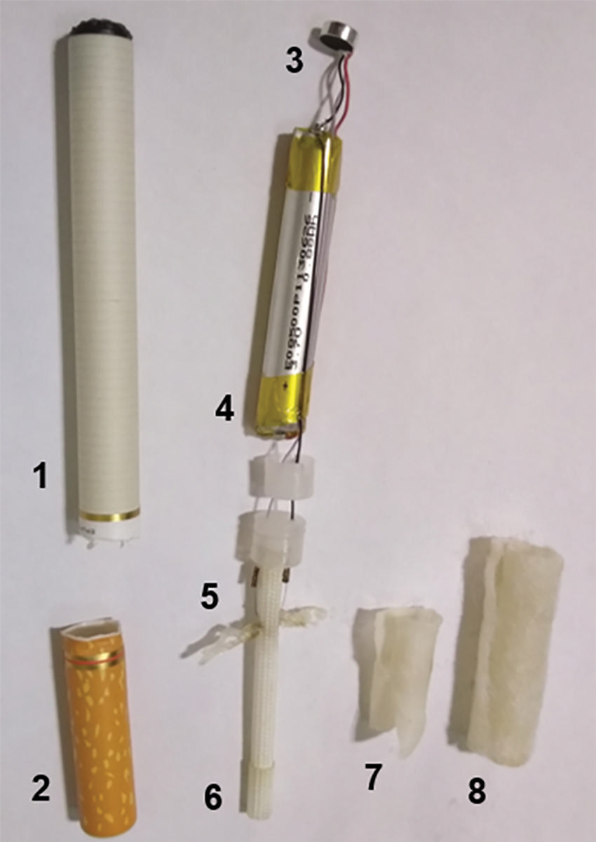
Figure 6. Electronic cigarette components. Photograph shows the superior envelope (1), mouthpiece (2), light emitting diode (3), cylindrical 3.7-V lithium battery (4), wick and filament (5), thick wire (6), inner fibers (7), and outer fibers (8).
High-res (TIF) version
(Right-click and Save As)

