Non-Contrast MRI is Effective in Monitoring MS Patients
Released: March 12, 2019
At A Glance
- MS patients can be effectively monitored without the use of contrast agents.
- Researchers assessed 507 follow-up MR images for new or enlarged lesions.
- The 3T MRI results did not differ significantly between contrast-enhanced and non-enhanced images.
- RSNA Media Relations
1-630-590-7762
media@rsna.org - Linda Brooks
1-630-590-7738
lbrooks@rsna.org - Dionna Arnold
1-630-590-7791
darnold@rsna.org
OAK BROOK, Ill. — Brain MRI without contrast agent is just as effective as the contrast-enhanced approach for monitoring disease progression in patients with multiple sclerosis (MS), according to a new study in the journal Radiology. The findings support the possibility that contrast enhancement can be omitted from routine follow-up scans.
MS is a disease in which the immune system attacks the body’s central nervous system, including the brain and spinal cord. This can lead to vision problems, numbness and a host of other symptoms. Damaged areas of the brain develop scar tissue, or lesions, that are visible on MRI.

MRI with the administration of gadolinium-based contrast material is widely considered obligatory for follow-up scans of patients with MS. Gadolinium, a heavy metal, enhances the images and helps provide important diagnostic information, but it leads to both prolonged scan times and increased costs. There is also evidence that some of the metal remains in the body after contrast administration, although the long-term clinical impact of these deposits is unclear.
“These factors warrant evaluation of strategies for reducing or omitting contrast agent, especially in MS patients who often accumulate a high number of MRI scans over their lifetimes,” said study senior author Benedikt Wiestler, M.D., from the Technische Universität München in Munich, Germany.
Advances in non-contrast MRI image acquisition and post-processing technology, along with the increasing availability of powerful 3T MRI machines, have raised the possibility that non-enhanced scans could have a role in MS follow-up imaging.
To learn more, Dr. Wiestler and colleagues used MRI to assess new or enlarged lesions in 359 patients with MS. Of 507 follow-up scans, 264 showed interval progression, defined by as at least one new or unequivocally enlarged lesion on follow-up MRI scans. There were a total of 1,992 new or enlarged lesions. With 3T MRI, the assessment of interval progression did not differ significantly between the contrast-enhanced and non-enhanced images.
“In over 500 follow-up scans, we missed only four of 1,992 new or enlarged lesions,” Dr. Wiestler said. “Importantly, we did not miss disease activity in the non-enhanced scans in a single follow-up scan.”
Dr. Wiestler credited an image subtraction pipeline developed and researched at his facility for the powerful sensitivity of the non-contrast MRI in detecting newly occurring lesions. The approach combines 3-D MRI and subtraction techniques, which cancel out unchanged areas in the follow-up image, substantially improving visualization of new or enlarging white matter lesions.
This combination of 3-D sequences and subtraction techniques is key to improving sensitivity for detecting newly occurred lesions, Dr. Wiestler said.
“Several vendors have made tools for generating subtraction images commercially available,” he said. “Implementing such tools into the routine clinical work flow will help to make the use of contrast agent dispensable in routine follow-up imaging of MS patients.”
“Accuracy of Unenhanced MRI in the Detection of New Brain Lesions in Multiple Sclerosis.” Collaborating with Dr. Wiestler were Paul Eichinger, M.D., Simon Schön, M.D., Viola Pongratz, M.D., Hanni Wiestler, M.D., Haike Zhang, Matthias Bussas, M.Sc., Muna-Miriam Hoshi, M.D., Jan Kirschke, M.D., Achim Berthele, M.D., Claus Zimmer, M.D., Bernhard Hemmer, M.D., and Mark Mühlau, M.D.
Radiology is edited by David A. Bluemke, M.D., Ph.D., University of Wisconsin School of Medicine and Public Health, Madison, Wis., and owned and published by the Radiological Society of North America, Inc. (https://pubs.rsna.org/journal/radiology)
RSNA is an association of over 53,400 radiologists, radiation oncologists, medical physicists and related scientists, promoting excellence in patient care and health care delivery through education, research and technologic innovation. The Society is based in Oak Brook, Ill. (RSNA.org)
For patient-friendly information on brain MRI RadiologyInfo.org.
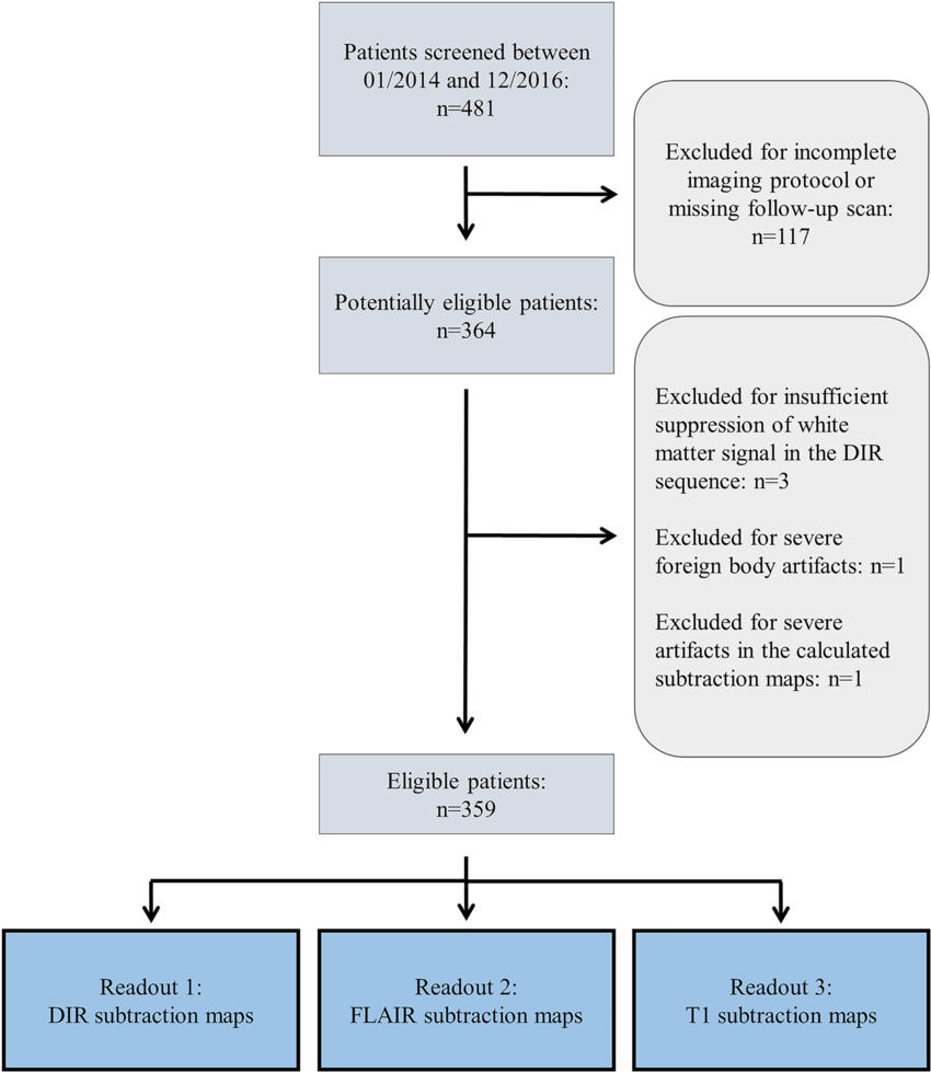
Figure 1. Flowchart shows inclusion and exclusion criteria. DIR = double inversion recovery, FLAIR = fluid attenuated inversion recovery.
High-res (TIF) version
(Right-click and Save As)
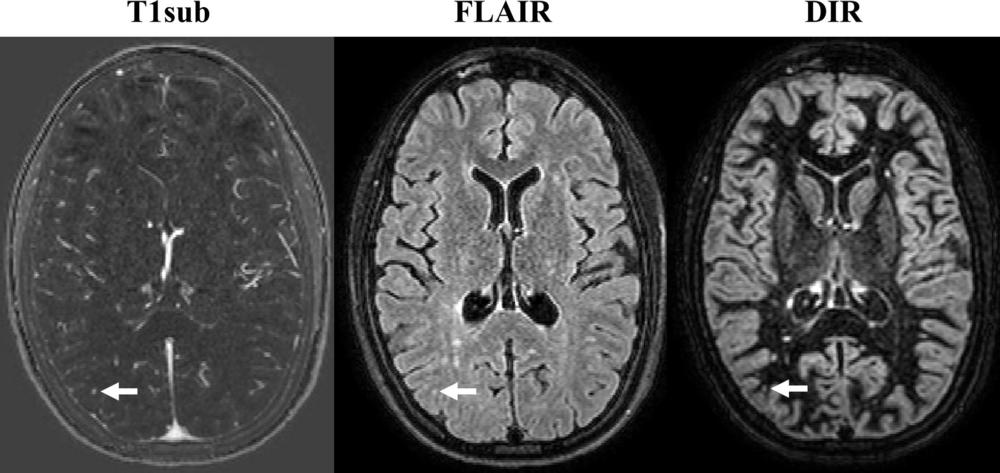
Figure 2. Axial MR images obtained in 32-year-old woman with relapsing-remitting multiple sclerosis. Images were obtained with subtraction of unenhanced T1-weighted MR image from contrast-enhanced MR image (T1sub), fluid-attenuated inversion recovery (FLAIR), and double inversion recovery (DIR). The new lesion (arrow), a small, subcortical lesion in right parietal lobe, is seen only on contrast-enhanced image; it was overlooked on DIR and FLAIR images. Note that there are several other new or enlarged lesions that can be seen on nonenhanced images.
High-res (TIF) version
(Right-click and Save As)
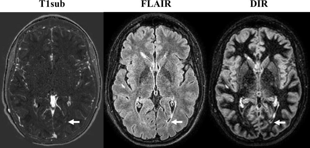
Figure 3. Axial MR images in 27-year-old man with relapsing-remitting multiple sclerosis. Images were obtained with subtraction of unenhanced T1-weighted MR image from contrast-enhanced MR image (T1sub), fluid-attenuated inversion recovery (FLAIR), and double inversion recovery (DIR). The new lesion (arrow), a small left periventricular lesion, is seen only on contrast-enhanced image; it was overlooked on DIR and FLAIR images. Note that there are several other new or enlarged lesions that are readily seen on nonenhanced images.
High-res (TIF) version
(Right-click and Save As)
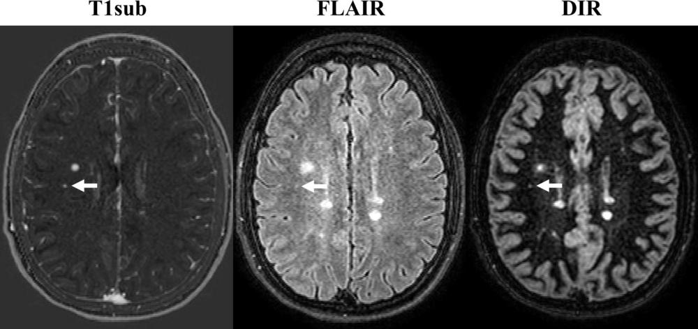
Figure 4. Axial MR images in 24-year-old woman with relapsing-remitting multiple sclerosis. Images were obtained with subtraction of unenhanced T1-weighted MR image from contrast-enhanced MR image (T1sub), fluid-attenuated inversion recovery (FLAIR), and double inversion recovery (DIR). The new lesion, a small subcortical lesion in right frontal lobe, was detected only on contrast-enhanced image; it was overlooked on DIR and FLAIR images. Note that there are several other new or enlarged lesions readily seen on nonenhanced images.
High-res (TIF) version
(Right-click and Save As)