Neurofeedback Shows Promise in Treating Tinnitus
Released: November 27, 2017
At A Glance
- Researchers are studying a novel approach to treat tinnitus, or "ringing in the ear."
- Using fMRI and neurofeedback, the researchers were able to train people to decrease brain activity in response to white noise.
- Tinnitus is a common ailment, affecting one in five people.
- RSNA Media Relations
1-630-590-7762
media@rsna.org - Maureen Morley
1-630-590-7754
mmorley@rsna.org - Linda Brooks
1-630-590-7738
lbrooks@rsna.org
CHICAGO — Researchers using functional MRI (fMRI) have found that neurofeedback training has the potential to reduce the severity of tinnitus or even eliminate it, according to a study presented today at the annual meeting of the Radiological Society of North America (RSNA).
Tinnitus is the perception of noise, often ringing, in the ear. The condition is very common, affecting approximately one in five people. As sufferers start to focus on it more, they become more frustrated and anxious, which in turn makes the noise seem worse. The primary auditory cortex, the part of the brain where auditory input is processed, has been implicated in tinnitus-related distress.
For the study, researchers looked at a novel potential way to treat tinnitus by having people use neurofeedback training to turn their focus away from the sounds in their ears. Neurofeedback is a way of training the brain by allowing an individual to view some type of external indicator of brain activity and attempt to exert control over it.
"The idea is that in people with tinnitus there is an over-attention drawn to the auditory cortex, making it more active than in a healthy person," said Matthew S. Sherwood, Ph.D., research engineer and adjunct faculty in the Department of Biomedical, Industrial and Human Factors Engineering at Wright State University in Fairborn, Ohio. "Our hope is that tinnitus sufferers could use neurofeedback to divert attention away from their tinnitus and possibly make it go away."
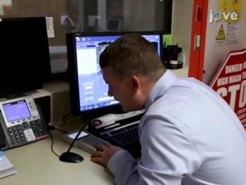
To determine the potential efficacy of this approach, the researchers had 18 healthy volunteers with normal hearing undergo five fMRI-neurofeedback training sessions. Study participants were given earplugs through which white noise could be introduced for periods of time. The earplugs also served to block out the scanner noise.
To obtain fMRI results, the researchers used single-shot echoplanar imaging, an MRI technique that is sensitive to blood oxygen levels, providing an indirect measure of brain activity.
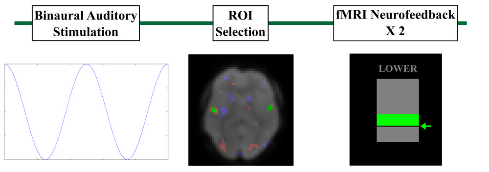
"We started with alternating periods of sound and no sound in order to create a map of the brain and find areas that produced the highest activity during the sound phase," Dr. Sherwood said. "Then we selected the voxels that were heavily activated when sound was being played."
The participants then participated in the fMRI-neurofeedback training phase while inside the MRI scanner. They received white noise through their earplugs and were able to view the activity in their primary auditory cortex as a bar on a screen. Each fMRI-neurofeedback training run contained eight blocks separated into a 30-second "relax" period followed by a 30-second "lower" period. Participants were instructed to watch the bar during the relax period and actively attempt to lower it by decreasing primary auditory cortex activity during the lower phase.
The researchers gave the participants techniques to help them do this, such as trying to divert attention from sound to other sensations like touch and sight.
"Many focused on breathing because it gave them a feeling of control," Dr. Sherwood said. "By diverting their attention away from sound, the participants' auditory cortex activity went down, and the signal we were measuring also went down."
A control group of nine individuals were provided sham neurofeedback — they performed the same tasks as the other group, but the feedback came not from them but from a random participant. By performing the exact same procedures with both groups using either real or sham neurofeedback, the researchers were able to distinguish the effect of real neurofeedback on control of the primary auditory cortex.
The study represents the first time fMRI-neurofeedback training has been applied to demonstrate that there is a significant relationship between control of the primary auditory cortex and attentional processes. This is important to therapeutic development, Sherwood said, as the neural mechanisms of tinnitus are unknown but likely related to attention.
The results represent a promising avenue of research that could lead to improvements in other areas of health like pain management, according to Dr. Sherwood.
"Ultimately, we'd like take what we learned from MRI and develop a neurofeedback program that doesn't require MRI to use, such as an app or home-based therapy that could apply to tinnitus and other conditions," he said.
Co-authors are Emily E. Diller, M.S., Subhashini Ganapathy, Ph.D., Jeremy Nelson, Ph.D., and Jason G. Parker, Ph.D. This material is based on research sponsored by the U.S. Air Force under agreement number FA8650-16-2-6702. The views expressed are those of the authors and do not reflect the official views or policy of the Department of Defense and its Components. The U.S. Government is authorized to reproduce and distribute reprints for Governmental purposes notwithstanding any copyright notation thereon. The voluntary, fully informed consent of the subjects used in this research was obtained as required by 32 CFR 219 and DODI 3216.02_AFI 40-402.
Note: Copies of RSNA 2017 news releases and electronic images will be available online at RSNA.org/press17 beginning Monday, Nov. 27.
RSNA is an association of over 54,000 radiologists, radiation oncologists, medical physicists and related scientists, promoting excellence in patient care and health care delivery through education, research and technologic innovation. The Society is based in Oak Brook, Ill. (RSNA.org)
Editor’s note: The data in these releases may differ from those in the published abstract and those actually presented at the meeting, as researchers continue to update their data right up until the meeting. To ensure you are using the most up-to-date information, please call the RSNA Newsroom at 1-312-791-6610.
For patient-friendly information on fMRI, visit RadiologyInfo.org.
Video (.mp4 format)
Video 1. Matthew Sherwood, Ph.D., and colleagues discussing the implementation of fMRI neurofeedback.
Download.mp4
(Right-click and Save As)
Video 2 Dr. Matthew S. Sherwood discusses his research on neurofeedback and tinnitus.
Download.mp4
(Right-click and Save As)
Video 3. Dr. Matthew S. Sherwood discusses the most important finding of his study.
Download.mp4
(Right-click and Save As)
Video 4. Dr. Matthew S. Sherwood discusses why he wanted to pursue this research.
Download.mp4
(Right-click and Save As)
Video 5. Dr. Matthew S. Sherwood explains what neurofeedback is.
Download.mp4
(Right-click and Save As)
Video 6. Dr. Matthew S. Sherwood’s advice to patients with tinnitus.
Download.mp4
(Right-click and Save As)
Video 7. Dr. Matthew S. Sherwood discusses what the next steps in his research are.
Download.mp4
(Right-click and Save As)
Images (.JPG and .TIF format)
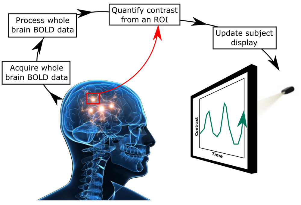
Figure 1. The standard approach to fMRI neurofeedback.
High-res (TIF) version
(Right-click and Save As)
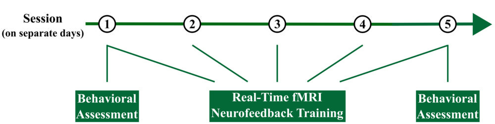
Figure 2. Overview of the experimental design. Each participant completed 5 sessions.
High-res (TIF) version
(Right-click and Save As)

Figure 4. Control over the primary auditory cortex (A1 control) separated by group and session. The experimental group was found to have significantly higher control, averaged across training, than the control group.
High-res (TIF) version
(Right-click and Save As)
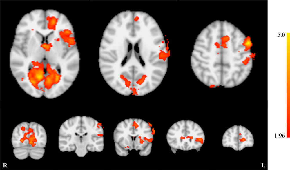
Figure 5. Whole brain effects of neurofeedback training.
High-res (TIF) version
(Right-click and Save As)
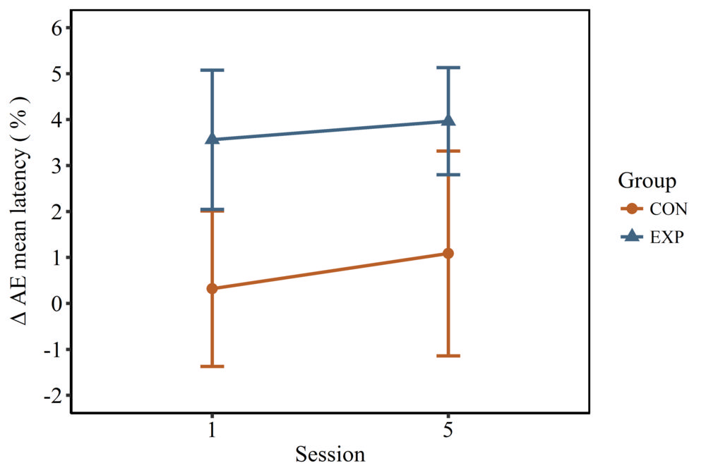
Figure 6. Effect of emotion on attention. Emotional distractors resulted in a significantly larger change in response latency in the experimental group when compared to the control group. However, the impact of emotion on attention was not found to change significantly between the groups across training.
High-res (TIF) version
(Right-click and Save As)
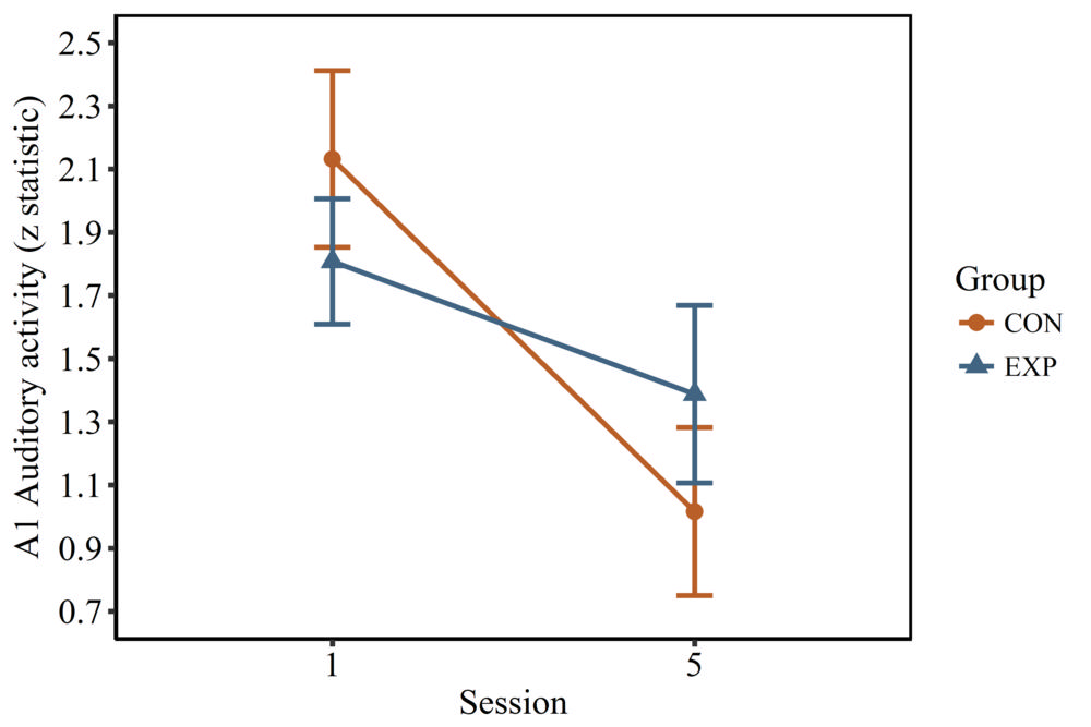
Figure 7. Activation of the primary auditory cortex in response to binaural stimulation. Activation significantly decreased from session 1 to session 5.
High-res (TIF) version
(Right-click and Save As)
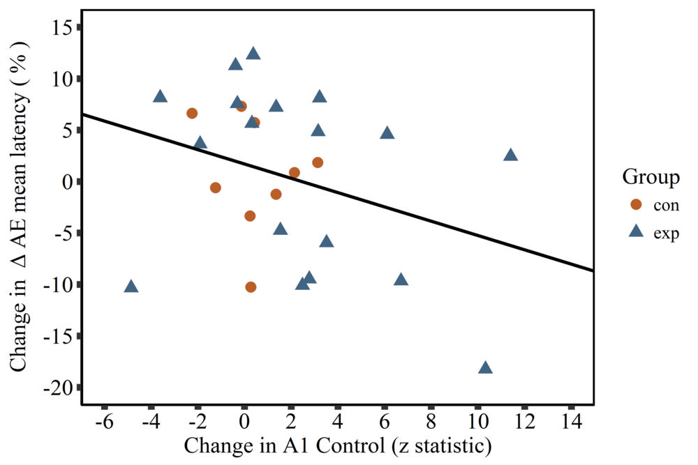
Figure 8. Improvements in control over the primary auditory cortex were found to be significantly related to decreases in the effect of emotion on attention.
High-res (TIF) version
(Right-click and Save As)