Study Looks at Needles in Treatment for Shoulder Pain
Released: June 14, 2017
At A Glance
- Ultrasound-guided percutaneous irrigation of calcific tendinopathy can be performed with one or two needles.
- Calcific tendinopathy is a condition in which calcium deposits form on the tendons of the rotator cuff, a group of four tendons that stabilizes the shoulder joint.
- Procedure times were shorter and the calcium dissolution was easier when using two needles for hard calcifications and one needle for fluid calcifications.
- RSNA Media Relations
1-630-590-7762
media@rsna.org - Linda Brooks
1-630-590-7738
lbrooks@rsna.org
OAK BROOK, Ill. — According to a new study published online in the journal Radiology, the type of procedure used to treat shoulder calcifications should be tailored to the type of calcification. The results of the study will help interventional radiologists determine whether to use one or two needles for an ultrasound-guided treatment for a common condition called rotator cuff calcific tendinopathy.

Calcific tendinopathy is a condition in which calcium deposits form on the tendons of the rotator cuff, a group of four tendons that stabilizes the shoulder joint. The condition, which occurs in approximately 20 percent of painful shoulders, causes pain and tenderness ranging from low-grade to highly disabling.
“There is still no consensus on how to treat calcific tendinopathy,” said researcher Luca Maria Sconfienza, M.D., Ph.D., associate professor at the University of Milan and chair of the Department of Radiology at the IRCCS Istituto Ortopedico Galeazzi. “However, ultrasound-guided percutaneous irrigation is widely performed throughout the world and is currently the first-line treatment for the condition, because it is quick, minimally invasive and has a low complication rate.”
The procedure involves injecting a fluid such as saline solution into the tendon to dissolve the calcium deposits and then extracting the calcium-filled solution.
“The main difference among ultrasound percutaneous irrigation procedures is the use of one or two needles,” Dr. Sconfienza said. “Until now, a direct comparison of one versus two needles has never been performed.”
The study included 211 patients (77 men and 134 women between the ages of 24 and 69) who underwent ultrasound percutaneous irrigation between 2012 and 2014. The patients were randomly assigned to have the one-needle or the two-needle procedure.
Ultrasound exams were performed on each patient to identify the exact location of calcium deposits in the tendons and whether the deposit appeared to be hard or fluid. For patients in the double-needle procedure group, 16-gauge needles were inserted inside the calcification under continuous ultrasound monitoring, and the area was flushed with injections and extractions of saline until the tendon was free of visible calcium. The single-needle procedure utilized an 18-gauge needle for the injection and extraction.
“Procedure times were shorter and the calcium dissolution was easier when using two needles for hard calcifications and one needle for fluid calcifications,” Dr. Sconfienza said.
“In terms of clinical outcomes after one year of follow-up, there was no significant difference between single- and double-needle ultrasound-guided irrigation,” he added.
At one-year follow-up, no residual or new calcifications or tendon tears at the site of the initial calcium deposits were detected.
“Rotator Cuff Calcific Tendinopathy: Randomized Comparison of US-guided Percutaneous Treatments by Using One or Two Needles.” Collaborating with Dr. Sconfienza were Davide Orlandi, M.D., Ph.D., Giovanni Mauri, M.D., Francesca Lacelli, M.D., Angelo Corazza, M.D., Carmelo Messina, M.D., Enzo Silvestri, M.D., and Giovanni Serafini, M.D.
Radiology is edited by Herbert Y. Kressel, M.D., Harvard Medical School, Boston, Mass., and owned and published by the Radiological Society of North America, Inc. (http://radiology.rsna.org/)
RSNA is an association of over 54,600 radiologists, radiation oncologists, medical physicists and related scientists, promoting excellence in patient care and health care delivery through education, research and technologic innovation. The Society is based in Oak Brook, Ill. (RSNA.org)
For patient-friendly information on interventional radiology, visit RadiologyInfo.org.
Images (.JPG and .TIF format)
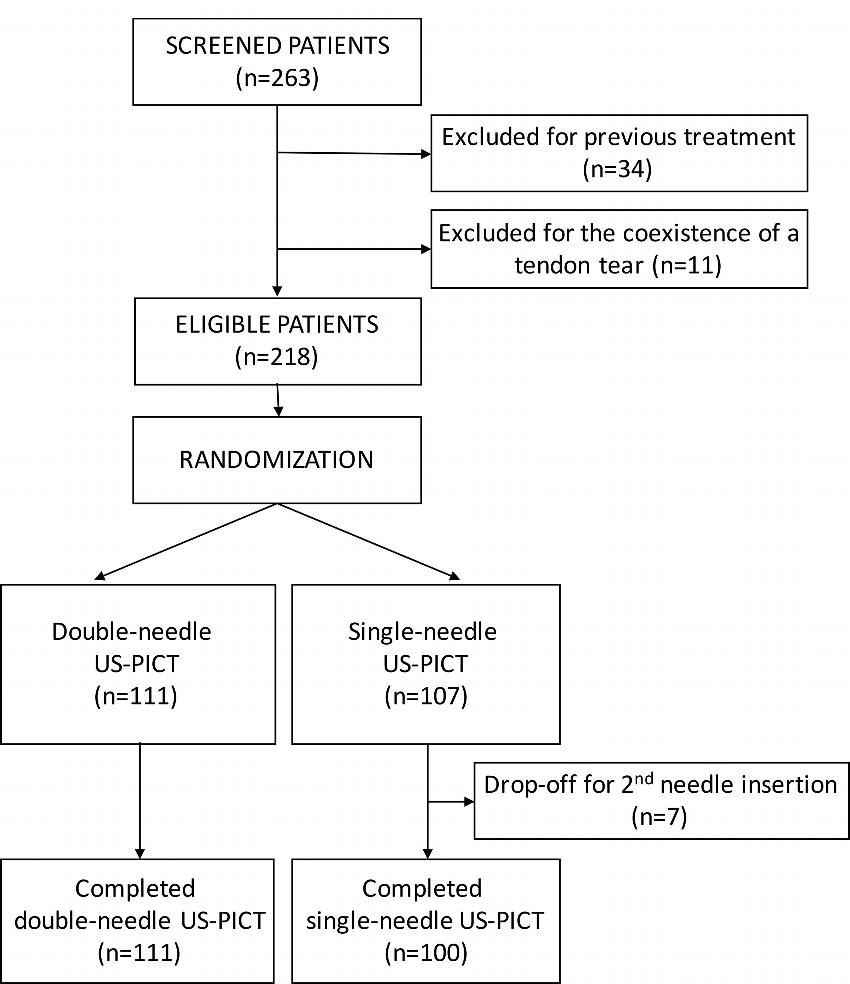
Figure 1. Flowchart of study population. US-PICT = ultrasonography-guided percutaneous irrigation of calcific tendinopathy.
High-res (TIF) version
(Right-click and Save As)
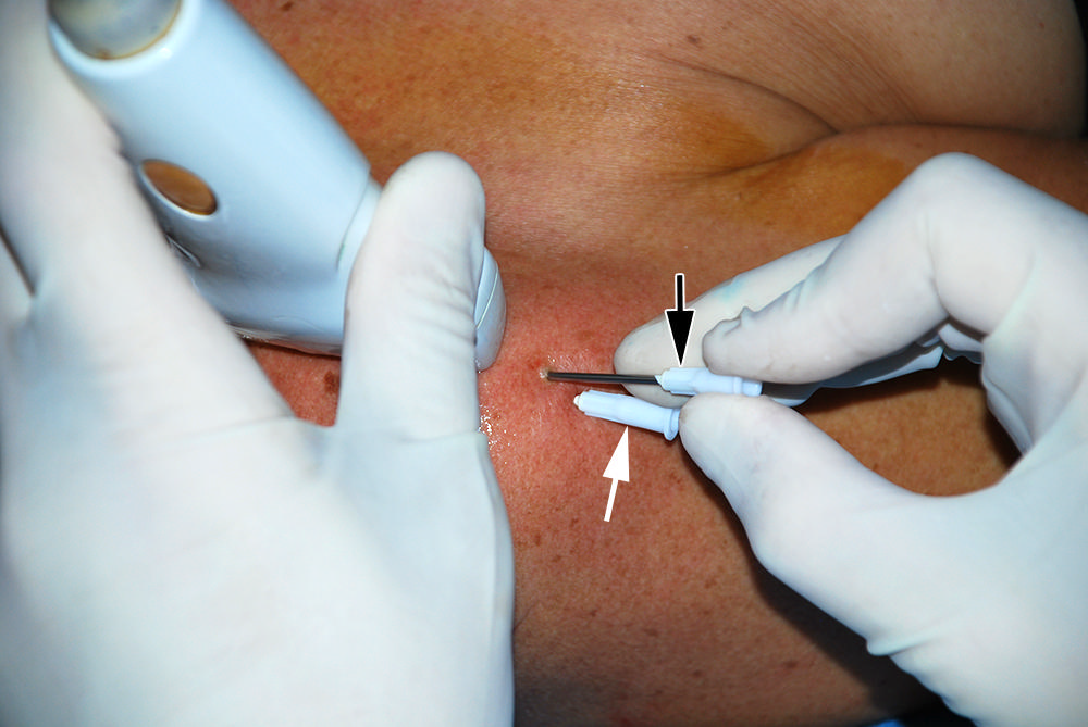
Figure 2. Photograph shows positioning of needles in a 56-year old woman treated with double-needle procedure. Needles are inserted from lateral side of shoulder. However, entrance position can be varied according to most convenient access to calcification. The more caudal needle (white arrow) should be inserted first to avoid image disturbance by the more cranial needle (black arrow). Needles should be inserted on same coronal plane, so they can be visualized together with a single scan during the procedure. When single-needle procedure is used, approach is the same but only the more caudal needle (white arrow) is used.
High-res (TIF) version
(Right-click and Save As)
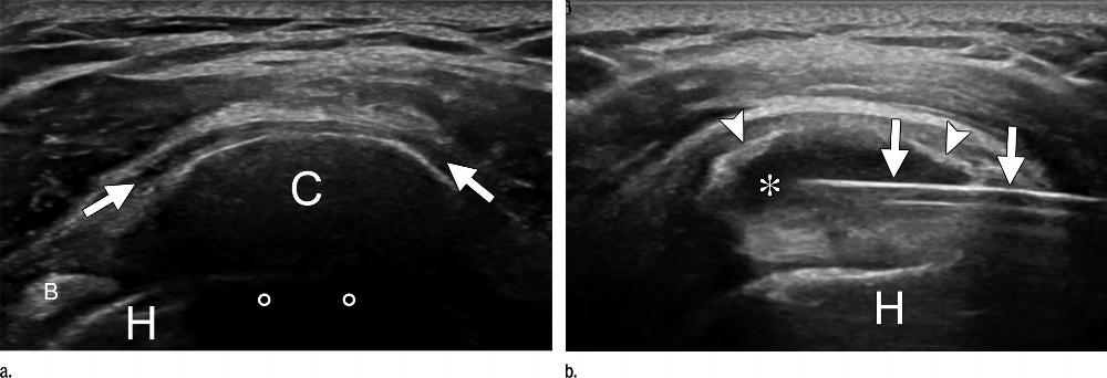
Figure 3. US images show supraspinatus calcification in a 29-year-old woman. (a) Calcific deposit (C) can be clearly seen with moderate acoustic shadowing (circles). Subacromial bursitis (arrows) can be seen. (b) End of treatment. Most calcific material was removed from calcification (∗) with single-needle procedure. Only a thin calcific wall (arrowheads) was left. Needle (arrows) is still inside calcification. B = biceps tendon, H = humeral head.
High-res (TIF) version
(Right-click and Save As)
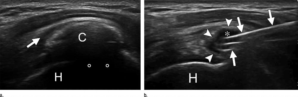
Figure 4. US images show supraspinatus calcification in a 37-year-old man. (a) Calcific deposit (C) can be clearly seen with moderate acoustic shadowing (circles). Subacromial bursitis (arrow) can be seen. (b) End of treatment. Most calcific material was removed from calcification (∗) with double-needle procedure. Only a thin calcific wall (arrowheads) was left. Needles (arrows) are still inside calcification. H = humeral head.
High-res (TIF) version
(Right-click and Save As)