Head Impacts Lead to Brain Changes in High School Football Players
Released: November 28, 2016
At A Glance
- Imaging exams found that high school football players without concussion showed structural and functional changes in the brain that correlated with exposure to head impacts.
- Players were imaged pre- and post-season, and wore special helmets that measured the magnitude, location and direction of each hit through one season.
- The findings showed that players who had greater head impact exposure had the greatest change in diffusion imaging and MEG metrics.
- RSNA Media Relations
1-630-590-7762
media@rsna.org - Maureen Morley
1-630-590-7754
mmorley@rsna.org - Linda Brooks
1-630-590-7738
lbrooks@rsna.org
CHICAGO — Brain imaging exams performed on high school football players after just one season revealed changes in both the gray and white matter that correlated with exposure to head impacts, according to a new study that will be presented today at the annual meeting of the Radiological Society of North America (RSNA).

"It's important to understand the potential changes occurring in the brain related to youth contact sports," said Elizabeth Moody Davenport, Ph.D., a postdoctoral researcher at UT Southwestern Medical Center in Dallas, Texas, who led this analysis. "We know that some professional football players suffer from a serious condition called chronic traumatic encephalopathy, or CTE. We are attempting to find out when and how that process starts, so that we can keep sports a healthy activity for millions of children and adolescents."
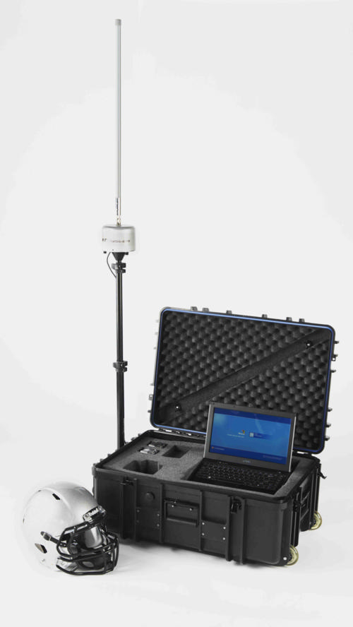
The study included 24 players from a high school football team in North Carolina, each of whom wore a helmet outfitted with the Head Impact Telemetry System (HITS) during all practices and games. The helmets are lined with six accelerometers, or sensors, that measure the magnitude, location and direction of a hit. Data from the helmets can be uploaded to a computer for analysis.
"We saw changes in these young players' brains on both structural and functional imaging after a single season of football," Dr. Davenport said.
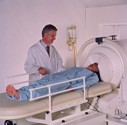
In the study, each player underwent pre- and post-season imaging: a specialized MRI scan, from which diffusion tensor imaging (DTI) and diffusion kurtosis imaging (DKI) data were extracted to measure the brain's white matter integrity, and a magnetoencephalography (MEG) scan, which records and analyzes the magnetic fields produced by brain waves. Diffusion imaging can measure the structural white matter changes in the brain, and MEG assesses changes in function.
"MEG can be used to measure delta waves in the brain, which are a type of distress signal," Dr. Davenport said. "Delta waves represent slow wave activity that increases after brain injuries. The delta waves we saw came from the surface of the brain, while diffusion imaging is a measure of the white matter deeper in the brain."
The research team calculated the change in imaging metrics between the pre- and post-season imaging exams. They measured abnormalities observed on diffusion imaging and abnormally increased delta wave activity on MEG. The imaging results were then combined with player-specific impact data from the HITS. None of the 24 players were diagnosed with a concussion during the study.
Players with greater head impact exposure had the greatest change in diffusion imaging and MEG metrics.
"Change in diffusion imaging metrics correlated most to linear acceleration, similar to the impact of a car crash," Dr. Davenport said. "MEG changes correlated most to rotational impact, similar to a boxer's punch. These results demonstrate that you need both imaging metrics to assess impact exposure because they correlate with very different biomechanical processes."
Dr. Davenport said similar studies are being conducted this fall, and a consortium has been formed to continue the brain imaging research in youth contact sports across the country.
"Without a larger population that is closely followed in a longitudinal study, it is difficult to know the long-term effects of these changes," she said. "We don't know if the brain's developmental trajectory is altered, or if the off-season time allows for the brain to return to normal."
Co-authors on the study are Jillian Urban, Ph.D., Ben Wagner, B.S., Mark A. Espeland, Ph.D., Christopher T. Whitlow, M.D., Ph.D., Joel Stitzel, Ph.D., and Joseph A. Maldjian, M.D.
Note: Copies of RSNA 2016 news releases and electronic images will be available online at RSNA.org/press16 beginning Monday, Nov. 28.
RSNA is an association of more than 54,000 radiologists, radiation oncologists, medical physicists and related scientists, promoting excellence in patient care and health care delivery through education, research and technologic innovation. The Society is based in Oak Brook, Ill. (RSNA.org)
Editor's note: The data in these releases may differ from those in the published abstract and those actually presented at the meeting, as researchers continue to update their data right up until the meeting. To ensure you are using the most up-to-date information, please call the RSNA Newsroom at 1-312-791-6610.
For patient-friendly information on brain MRI, visit RadiologyInfo.org.
Images (.JPG and .TIF format)
and Video clips (.mp4 format)
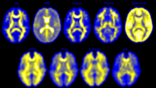
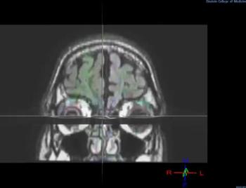
Figure 1. Diffusion Tensor Imaging (DTI) of the top part of the brain visible in the top row; Diffusional Kurtosis Imaging (DKI) of the bottom part of the brain visible in the bottom row.
High-res (TIF) version
(Right-click and Save As)
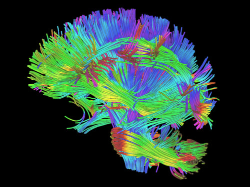
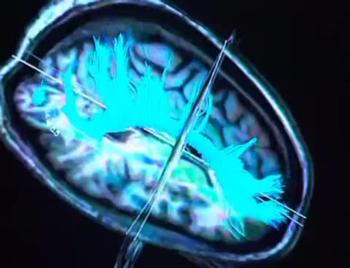
Figure 2. Example of a white matter rendering from DTI and DKI scans. The diffusion metrics used in this study were derived from DTI and DKI scans. The metrics give measures of the integrity of these white matter fibers.
High-res (TIF) version
(Right-click and Save As)

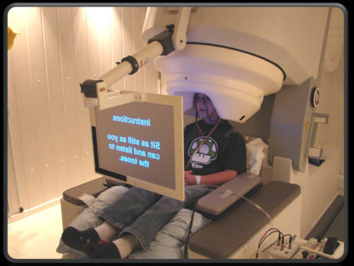
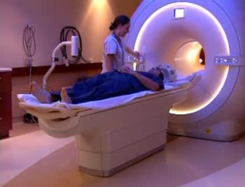
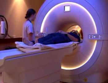
Figure 3. Magnetoencephalography (MEG) machine at Wake Forest Baptist Medical Center. The green dots represent the fit of the sensors around a subject’s brain.
High-res (TIF) version
(Right-click and Save As)
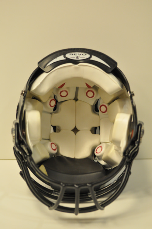
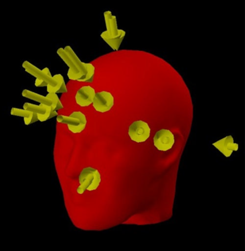
Figure 5. Vectors representing head impacts recorded with the Head Impact Telemetry (HIT) System.
High-res (TIF) version
(Right-click and Save As)
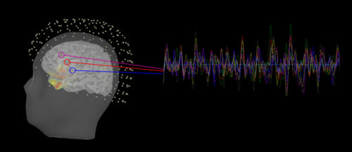
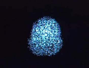
Figure 6. Delta waves in the brain of a subject after a season of high school football. The dots above the head represent the MEG sensors and the colored lines represent the time series signals. The highlighted portion of the brain represents an area with a high concentration of delta waves.
High-res (TIF) version
(Right-click and Save As)

Figure 7. Riddell HIT System with a sensor array and helmet
High-res (TIF) version
(Right-click and Save As)
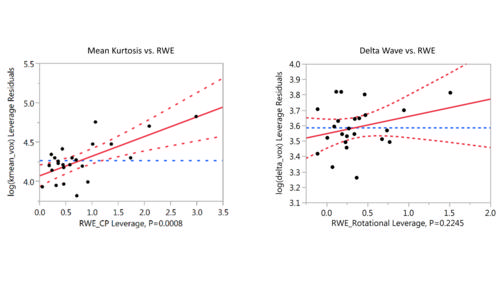
Figure 8. (Left) DKI metric, mean kurtosis, has previously shown a positive linear relationship with RWE; (Right) Increased delta waves have previously shown a positive linear relationship with RWE.
High-res (TIF) version
(Right-click and Save As)
Video 1. Animation showing how the HIT system accelerometers fit around the subject’s head. The springs ensure the sensors remain in contact with the head, measuring head acceleration not helmet acceleration.
Download.mp4
(Right-click and Save As)
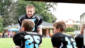
Video 2. B-roll footage of adolescents playing football using helmets equipped with HITS.
Download.mp4
(Right-click and Save As)


Video 9. Elizabeth Davenport, Ph.D., discusses the findings of her research on brain changes in high school football players.br>
Download.mp4
(Right-click and Save As)

Video 10. Elizabeth Davenport, Ph.D., discusses why she conducted the research on high school football players.
Download.mp4
(Right-click and Save As)

Video 11. Elizabeth Davenport, Ph.D., explains how this study is different from earlier studies.
Download.mp4
(Right-click and Save As)

Video 12. Elizabeth Davenport, Ph.D., explains why the research is important.
Download.mp4
(Right-click and Save As)

Video 13. Elizabeth Davenport, Ph.D., discusses concerns that parents may have about their teenagers playing football.
Download.mp4
(Right-click and Save As)