New Studies Provide More Insight into Zika Effects
Released: November 22, 2016
At A Glance
- A pattern of CT findings show that Zika virus causes several types of congenital brain damage.
- Some adults who contract Zika virus develop serious neurological conditions, such as Guillain-Barré syndrome.
- Knowledge of abnormalities present in the central nervous system due to Zika infection may give hints about the pathophysiology of the disease.
- RSNA Media Relations
1-630-590-7762
media@rsna.org - Maureen Morley
1-630-590-7754
mmorley@rsna.org - Linda Brooks
1-630-590-7738
lbrooks@rsna.org
CHICAGO — Three new studies reporting on the effects of the Zika virus outbreak in Brazil will be presented next week at the annual meeting of the Radiological Society of North America (RSNA).
The first study looks at CT findings of the central nervous system in 16 newborn babies with congenital Zika virus infection confirmed by tests in cerebral spinal fluid.
The researchers identified a pattern of CT brain findings in the babies, including decreased brain volume, simplified gyral pattern, calcifications, ventricular dilatation and prominent occipital bone.

"We live in Pernambuco, a state in northeastern Brazil, which had the highest number of patients with microcephaly during the Zika outbreak in our country," said study author Natacha Calheiros de Lima Petribu, M.D., from the Department of Radiology at Barão de Lucena Hospital. "Our study proves that Zika virus infection can cause congenital brain damage in babies with and without microcephaly."
Another study analyzed the imaging results of three target groups affected by Zika: adults who developed acute neurological syndrome, newborns with vertical infection with neurological disorders, and pregnant women with rash outbreaks suggestive of Zika.
Many of the adults had symptoms of Guillain-Barré syndrome, a rare disorder in which the body's immune system attacks the nervous system causing rapid onset muscle weakness. A few showed inflammation of the brain and spinal cord (Bickerstaff's encephalitis) or brain stem and spinal cord lesions. Common MRI findings included enhancement of certain spinal and facial nerves. In the newborns, MRI showed orbital injuries and anatomical changes in brain tissue.

"It was alarming to find so many cases of neurological syndromes in adults, some very serious, related to Zika virus infection," said study author Emerson de Melo Casagrande, M.D., from the Department of Radiology at Antonio Pedro University Hospital - Federal Fluminense University. "We have also noticed a difference between these syndromes, even though the trigger was the same."
In a third study, ultrasound and fetal MRI were performed on pregnant patients with Zika virus infection at different gestational ages. Once the babies were born, they underwent ultrasound, CT and MRI. The researchers then created 3-D virtual and physical models of the skulls. More than half the babies had microcephaly, brain calcifications and loss of brain tissue volume, along with other structural changes.
"The emergence of Zika virus in the Americas has coincided with increased reports of babies born with microcephaly," said study author Heron Werner Jr., M.D., Ph.D., from the Department of Radiology at Clínica de Diagnóstico por Imagem. "An early diagnosis may help in treating these babies after birth. Moreover, the knowledge of abnormalities present in the central nervous system may give hints about the pathophysiology of the disease."

Zika is mainly spread to humans via the bite of an infected mosquito. Symptoms may include fever, rash, joint or muscle pain, headache and bloodshot eyes. More serious conditions, such as Guillain-Barré syndrome, have been associated with Zika infection in adults, but are uncommon. Many adults infected with Zika have no symptoms at all.
Zika appears to be most dangerous when transmitted from a pregnant mother to her fetus during the first trimester of pregnancy, increasing the likelihood of severe brain defects in the baby, including microcephaly. Zika has also been linked to eye defects, hearing impairment and stunted growth in babies.
Pregnant women and women who are considering becoming pregnant should avoid visiting areas where infected mosquitos are known to be present. However, if women live in areas where the mosquitos are present, the recommendation of the Centers for Disease Control and Prevention (CDC) is to wear clothes that protect from mosquito bites, use mosquito repellent and get appropriate testing, including routine prenatal care and an ultrasound at 18 to 20 weeks. Pregnant women who are worried that they may have contracted the virus should speak with their obstetrician to initiate testing.
Although these reports focus on the Zika outbreak in Brazil, the infection has spread to countries and territories around the world, including the United States.
"Common Findings on Head Computed Tomography in Neonates with Confirmed Congenital Zika Syndrome": Natacha Calheiros de Lima Petribu, M.D., Marilia B. Abath, M.D., Andrezza C. Vieira Fernandes, M.D., Felipe R. Queiroz, M.D., Jannie D. Araujo, M.D., Glauber B. Carvalho, M.D., and Vanessa van der Linden, M.D.
"Neuroradiological Findings Related to Zika Epidemic:Experience from a Brazilian University Hospital": Cristina A. Fontes, M.D., M.Mc., Alair Augusto S. Santos, M.D., Ph.D., Emerson de Melo Casagrande, M.D., Victor M. Bussed, M.D., Daniel G. Neves, M.D., and Alessandro S. Melo, M.D., Ph.D.
"Essentials of Intrauterine Zika Virus Infection: Pre and Postnatal CNS Findings": Bianca Guedes Ribeiro, M.D., Heron Werner, M.D., Luiz Celso H. Da Cruz, M.D., Pedro Daltro, M.D., Renata A. Nogueira, M.D., and Tatiana M. Fazecas, M.D.
Note: Copies of RSNA 2016 news releases and electronic images will be available online at RSNA.org/press16 beginning Monday, Nov. 28.
RSNA is an association of more than 54,000 radiologists, radiation oncologists, medical physicists and related scientists, promoting excellence in patient care and health care delivery through education, research and technologic innovation. The Society is based in Oak Brook, Ill. (RSNA.org)
Editor's note: The data in these releases may differ from those in the published abstract and those actually presented at the meeting, as researchers continue to update their data right up until the meeting. To ensure you are using the most up-to-date information, please call the RSNA Newsroom at 1-312-791-6610.
For patient-friendly information on CT, MRI and ultrasound, visit RadiologyInfo.org.
Images (.JPG and .TIF format)
and Video clips (.mp4 format)
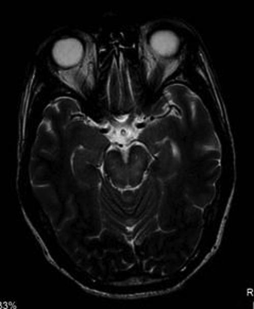
Figure 1. Cross section of an adult head where change is evident in the midbrain, being part of Bickerstaff encephalitis. This central structure is where cross neurons that control the movements of the limbs, and other organs such as the eyes, and controls the patient's consciousness. Changes to this area cause paralysis and coma. This change does not enhance with contrast agent (gadolinium), having a different pathophysiology from that seen in Guillain-Barré syndrome.
High-res (TIF) version
(Right-click and Save As)
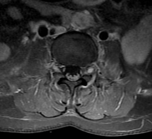
Figure 2. The lumbar spine, in cross-sectional view, where the spinal nerves enhance with intravenous contrast (gadolinium), demonstrating the inflammation that occurs in Guillain-Barré syndrome, occurring loss of limb movements. There is also observed enhancement in the spinal ganglion, shining next to the spine corresponding to the sensory changes that patients present.
High-res (TIF) version
(Right-click and Save As)
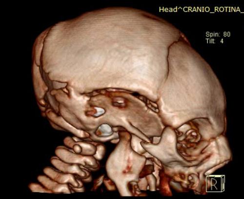
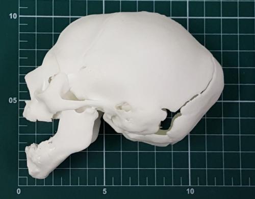
Figure 3. Skull 3D reconstruction of a newborn with microcephaly. Early fusion of the skull bones are observed, coursing with microcephaly and a cyclist helmet format seen in many of these children.
High-res (TIF) version
(Right-click and Save As)
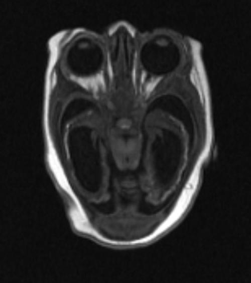
Figure 4. Angled cross image of a newborn, where significant thinning of brain tissue is found, with absence of cortical gyri and presence of calcifications inside. Dilation of cerebral ventricles are also observed.
High-res (TIF) version
(Right-click and Save As)
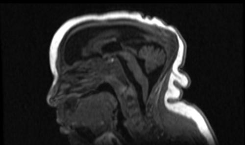
Figure 5. Side view of the same newborn (Picture 4) on the line between the cerebral hemispheres, showing an undeveloped corpus callosum, which is the structure that connects the two hemispheres of the brain, as well as enlargement of the cerebral fluid space. Microcephaly with the cyclist helmet skull format with excess of redundant skin on the neck is also shown.
High-res (TIF) version
(Right-click and Save As)
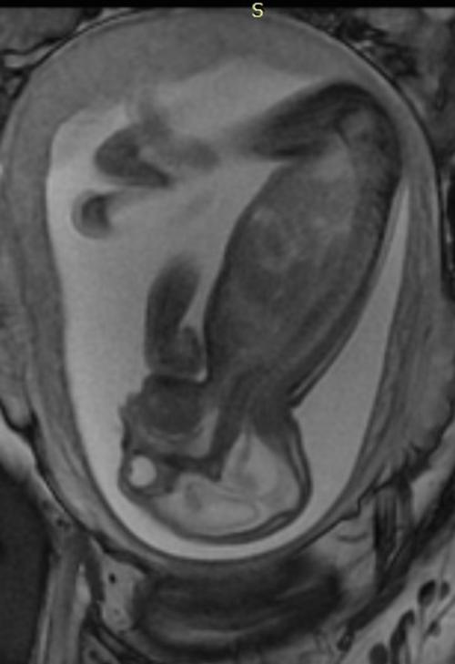
Figure 6. Side view of a fetus with hands and clubfeet (arthrogryposis), enlargement of the cerebral fluid space and dilation of cerebral ventricles. Thinning of the brain tissue, a poorly developed cerebellum and the absence of brain cortical gyri.
High-res (TIF) version
(Right-click and Save As)
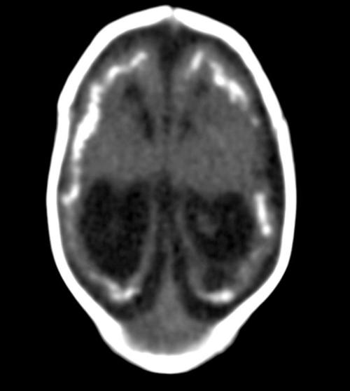
Figure 7. Unenhanced head CT showing brain atrophy, calcifications in cortico-subcortical junction, simplified gyral pattern and ventricular dilatation in a neonate with Congenital Zika Syndrome.
High-res (TIF) version
(Right-click and Save As)
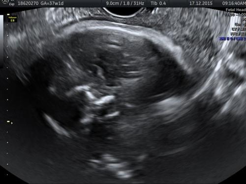
Figure 8. Prenatal ultrasound showing calcifications and microcephaly.
High-res (TIF) version
(Right-click and Save As)
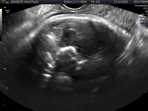
Figure 9. Prenatal ultrasound showing calcifications and microcephaly.
High-res (TIF) version
(Right-click and Save As)

Figure 10. Prenatal ultrasound showing calcifications and microcephaly.
High-res (TIF) version
(Right-click and Save As)
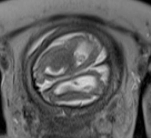
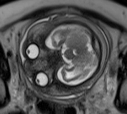
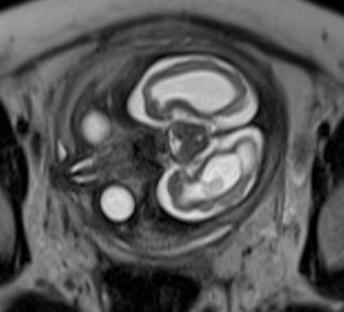
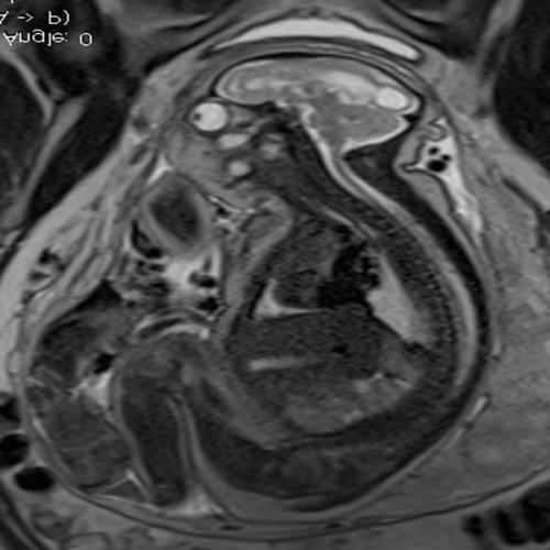
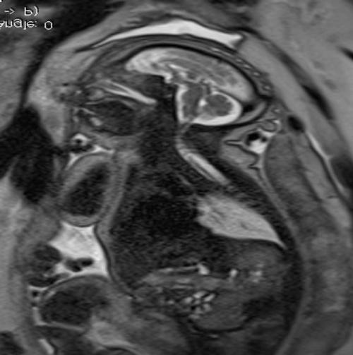
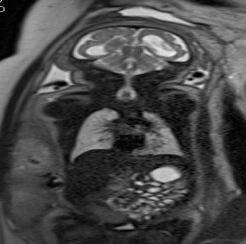

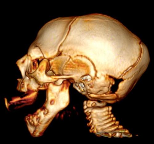
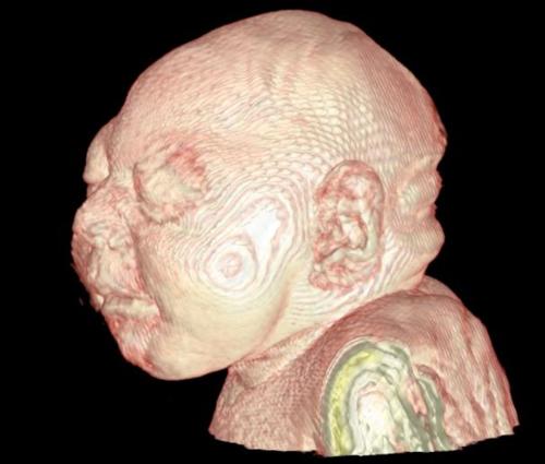
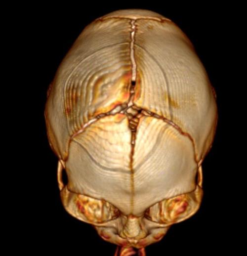
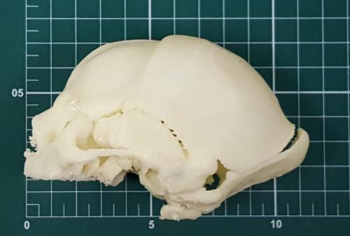
Figure 20. 3-D bone reconstruction from CT. The anterior fontanelle is small and the cranial sutures are tapered, but still pervious.
High-res (TIF) version
(Right-click and Save As)
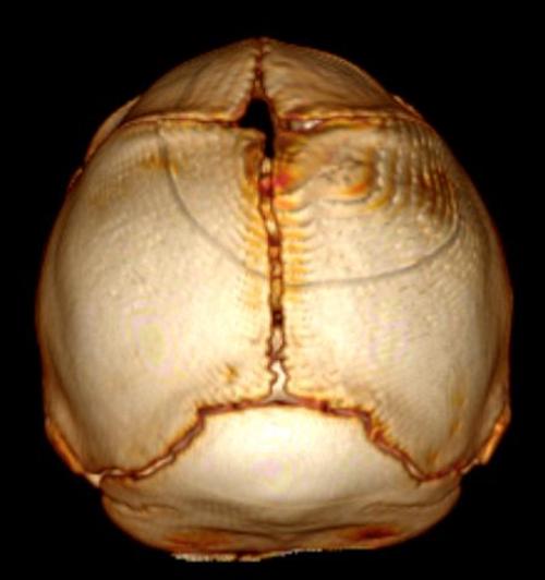
Figure 21. 3-D bone reconstruction from CT. The anterior fontanelle is small and the cranial sutures are tapered, but still pervious.
High-res (TIF) version
(Right-click and Save As)
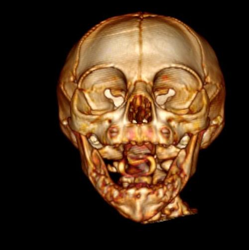
Figure 22. 3-D bone reconstruction from CT. The anterior fontanelle is small and the cranial sutures are tapered, but still pervious.
High-res (TIF) version
(Right-click and Save As)

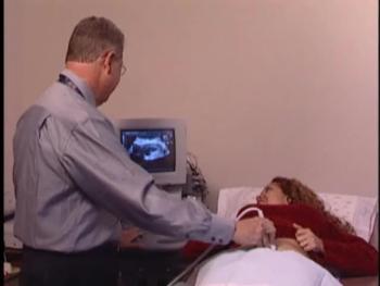
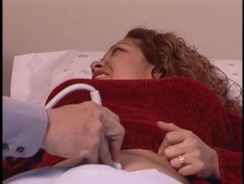
Video 1. A radiologist performs a Doppler ultrasound procedure on a pregnant patient.
Download.mp4
(Right-click and Save As)
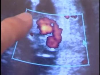
Video 3. The color seen here on the display represents pulsating blood of the fetal heart.
Download.mp4
(Right-click and Save As)

Video 4. Protection From Zika - Natacha Calheiros de Lima Petribu, M.D., discusses how to protect yourself from the Zika virus.
Download.mp4
(Right-click and Save As)

Video 5. Surprising Zika Findings - Natacha Calheiros de Lima Petribu, M.D., discusses Zika and the surprising findings of her research on infants.
Download.mp4
(Right-click and Save As)

Video 6. Interest In Zika Research - Marilia B. Abath, M.D., discusses why her team was interested in researching the effects of ZIka virus in infants.
Download.mp4
(Right-click and Save As)

Video 7. Findings of Zika Research - Marilia B. Abath, M.D., discusses the findings of her research of the ZIka virus in infants.
Download.mp4
(Right-click and Save As)

Video 8. Zika Research - Marilia B. Abath, M.D., talks about her Zika research.
Download.mp4
(Right-click and Save As)

Video 9. Zika Research - Heron Werner Jr., M.D., Ph.D., gives an overview of his Zika virus study.
Download.mp4
(Right-click and Save As)

Video 10. Zika Research - Heron Werner Jr., M.D., Ph.D., describes the findings of his study.
Download.mp4
(Right-click and Save As)

Video 11. Zika Research - Heron Werner Jr., M.D., Ph.D., describes surprising findings of his study.
Download.mp4
(Right-click and Save As)

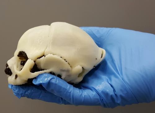
Video 12. Zika Research - Heron Werner Jr., M.D., Ph.D., uses a 3-D-printed skull model to show Zika effects in microcephaly.
Download.mp4
(Right-click and Save As)