‘Traffic Jam’ in Brain Linked to Common Cognitive Disorder
Released: September 06, 2016
At A Glance
- Brain MRI could help improve the diagnosis of people with vascular cognitive disorder.
- Researchers assessed brain MRI and cognitive examination results from 108 patients with symptomatic carotid artery disease, a risk factor for vascular cognitive disorder.
- The results point to disruption in the brain’s communication networks as a key mechanism of vascular cognitive disorder.
- RSNA Media Relations
1-630-590-7762
media@rsna.org - Linda Brooks
1-630-590-7738
lbrooks@rsna.org
OAK BROOK, Ill. — Brain MRI could help improve the diagnosis of people with a common type of cognitive disorder, according to a new study published online in the journal Radiology.
Vascular cognitive disorder is caused by disease of the vessels supplying blood to the brain. Strokes and transient ischemic attacks, or ministrokes, are risk factors. The resulting loss of healthy brain tissue adversely affects concentration and decision making and leads to problems with planning and organizing. Prevalence of vascular cognitive disorder is increasing in the elderly, and it can be challenging to diagnose and differentiate from other forms of dementia like Alzheimer's disease.

"If vascular cognitive disorder follows a major stroke, the cognitive impairment typically develops suddenly, and can thus be well recognized," said study co-author Dewen Meng, M.Sc., from the University of Nottingham in Nottingham, England. "In the majority of cases such a clear association is lacking, explaining why the detection of vascular cognitive disorder remains challenging."
A biomarker that could predict and track vascular cognitive disorder on imaging exams has yet to be found, although MRI measurements of the brain's signal-carrying white matter are a promising area of research. Damage to the white matter can be assessed with diffusion tensor imaging, an MRI technique that provides two important measures of microscopic brain damage: mean diffusivity and fractional anisotropy. Increased mean diffusivity, which measures the movement of water through tissue, is particularly sensitive to the breakdown of nerve fibers in the brain and the protective coating around them.
For the new study, researchers assessed brain MRI and cognitive examination results from 108 patients with symptomatic carotid artery disease, a risk factor for vascular cognitive disorder. Of the 108 patients, 53 were cognitively impaired. Further analysis showed a clear correlation between cognitive performance and the presence of chronic vascular disease-related lesions within certain white matter tracts of the brain. White matter tract skeleton mean diffusivity showed the closest correlation with impaired cognitive performance, making it a promising tool for improved diagnostic accuracy of vascular cognitive disorder.
"Using standard clinical brain MRI, we found that microscopic damage of main white matter tracts allowed us to distinguish patients with symptomatic carotid artery disease and cognitive impairment from those who were cognitively intact," said the study's senior author, Dorothee P. Auer, Ph.D., from the University of Nottingham. "Our findings mean that a simple MRI test might improve the diagnostic work-up of people with suspected vascular cognitive disorder, and holds further promise to track progression of the disorder."
The results suggest that a key mechanism of vascular cognitive disorder is subcortical disconnection, a kind of breakdown of communication within the large-scale cognitive neural networks.
Dr. Auer explained that the brain is functionally organized into networks that require efficient communication across specific brain regions. This communication depends on the information flow between the network nodes, not unlike the flow of passenger traffic through an extensive urban subway system. Subcortical disconnection suggests a disruption in connections between such cognitive nodes, impairing information flow and thus network coordination.
"The analogy would be a construction site at one of the main subway lines causing traffic disruptions," Dr. Auer said.
The researchers plan to expand their studies and look at changes in patients over time to see if they can track the progression of subcortical disconnection.
"This will be a critical step in the quest for prevention of vascular dementia, by helping to identify those at risk, and by enabling imaging studies to evaluate the effectiveness of interventions," Meng said.
The data used in the study is funded by National Institute for Health Research.
"Lesion Topography and Microscopic White Matter Tract Damage Contribute to Cognitive Impairment in Symptomatic Carotid Artery Disease." Collaborating with Meng and Dr. Auer were Akram A. Hosseini, Ph.D., Richard J. Simpson, Ph.D., Quratulain Shaikh, M.Sc., Christopher R. Tench, Ph.D., and Robert A. Dineen, Ph.D.
Radiology is edited by Herbert Y. Kressel, M.D., Harvard Medical School, Boston, Mass., and owned and published by the Radiological Society of North America, Inc. (http://radiology.rsna.org/)
RSNA is an association of more than 54,000 radiologists, radiation oncologists, medical physicists and related scientists, promoting excellence in patient care and health care delivery through education, research and technologic innovation. The Society is based in Oak Brook, Ill. (RSNA.org)
For patient-friendly information on MRI, visit RadiologyInfo.org.
Images (.JPG and .TIF format)
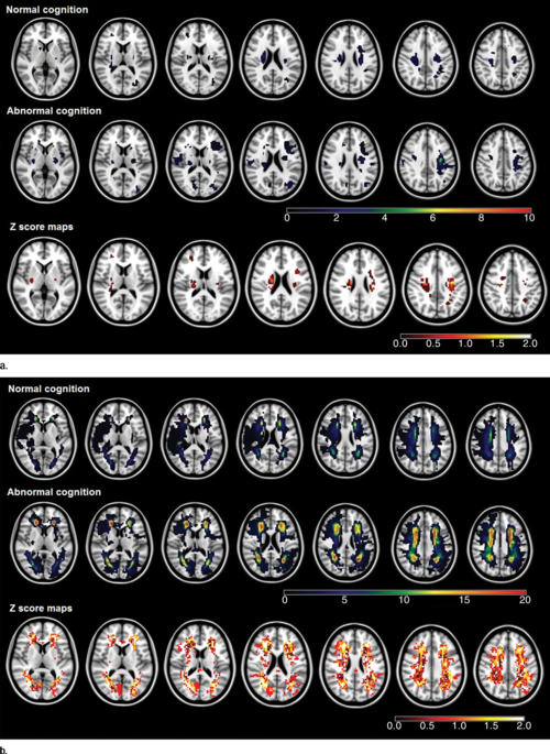
Figure 1. (a) Infarction maps in patients with normal cognition and those with abnormal cognition. Maps were superimposed onto the 1 X 1 X 1-mm MNI152 standard-space T1-weighted average structural template. The color bars show the variation between the minimum and maximum number of lesions. Z score maps represent the result of group comparison between patients with normal cognition and those with abnormal cognition. The significant voxels (according to results of the Fisher test) did not survive the false discovery rate correction. The color bar of the Z score maps shows the variation in Z score between the minimum and maximum values. (b) Chronic subcortical ischemic lesion frequency maps in patients with normal cognition and those with abnormal cognition. Details are the same as for a.
High-res (TIF) version
(Right-click and Save As)
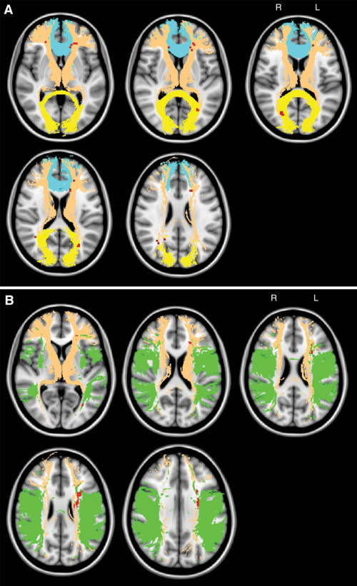
Figure 2. A, Maps show the topography of chronic ischemic lesions associated with global cognitive impairment. Significant clusters (red) ac¬cording to VLSM analysis (P ˂ .05, false discovery rate corrected, controlled for age and normalized total ischemic lesion volumes after logarithmic transformation) are projected onto the MNI152 standard-space T1-weighted average structural template image with overlain selected WMTs from the Johns Hopkins University DTI-based white matter atlas (yellow, forceps major; brown, anterior thalamic radiation; blue, forceps minor). B, Maps show the topography of chronic ischemic lesions associated with impaired fluency. Details are the same as for A (green, superior longitudinal fascicu¬lus). L = left, R = right.
High-res (TIF) version
(Right-click and Save As)
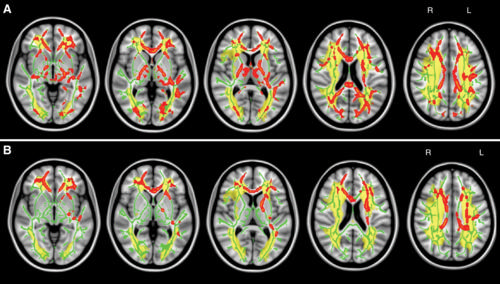
Figure 3. A, Maps show WMT mean diffusivity and global cognition. Significant TBSS results show skeletal voxels (red) where increased mean diffusivity correlated with global cognitive decline (controlled for age and normalized total ischemic lesion volumes after logarithmic transformation, P ˂ .05, corrected). Mean TBSS tract skeleton (green) and total ischemic lesion distribution map (yellow) were overlaid on the MNI152 standard-space T1-weighted average structural template image. B, Maps show WMT mean diffusivity and fluency. Details are the same as for A. L = left, R = right.
High-res (TIF) version
(Right-click and Save As)

Figure 4. Scatterplots show the correlation between mean diffusivity (MD) within chronic subcortical ischemic lesions, NAWM, WMT skeleton, and global cognitive performance, corrected for age.
High-res (TIF) version
(Right-click and Save As)
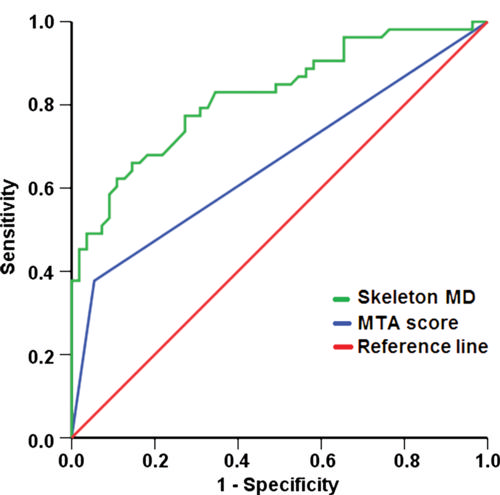
Figure 5. Receiver operating characteristic curves with a diagonal reference line (red) for prediction of global cognitive performance according to WMT skeleton mean diffusivity (MD, green) and MTA score (blue).
High-res (TIF) version
(Right-click and Save As)