Insomnia Linked to Damage in Brain Communication Networks
Released: April 05, 2016
At A Glance
- Twenty-three insomnia patients and 30 healthy controls were examined with diffusion tensor imaging and given questionnaires to evaluate mental status and sleep patterns.
- Compared to the healthy controls, insomnia patients had altered white matter tract integrity in brain regions that regulate sleep, consciousness, and alertness.
- White matter integrity abnormalities in insomnia patients may be caused by loss of myelin.
- RSNA Media Relations
1-630-590-7762
media@rsna.org - Linda Brooks
1-630-590-7738
lbrooks@rsna.org
OAK BROOK, Ill. — Using a sophisticated MRI technique, researchers have found abnormalities in the brain's white matter tracts in patients with insomnia. Results of the study were published online in the journal Radiology.
Primary insomnia, in which individuals have difficulty falling or staying asleep for a month or longer, is associated with daytime fatigue, mood disruption and cognitive impairment. Insomnia can also lead to depression and anxiety disorders.

"Insomnia is a remarkably prevalent disorder," said researcher Shumei Li, M.S., from the Department of Medical Imaging, Guangdong No. 2 Provincial People's Hospital, Guangzhou, China. "However, its causes and consequences remain elusive."
For the study, Li, along with colleagues lead by investigator Guihua Jiang, M.D., set out to analyze the white matter tracts in insomnia patients and the relationship between abnormal white matter integrity and the duration and features of insomnia.
"White matter tracts are bundles of axons—or long fibers of nerve cells—that connect one part of the brain to another," Li said. "If white matter tracts are impaired, communication between brain regions is disrupted."
The study included 23 patients with primary insomnia and 30 healthy control volunteers. To evaluate mental status and sleep patterns, all participants completed questionnaires including the Pittsburgh Sleep Quality Index, the Insomnia Severity Index, the Self-Rating Anxiety Scale and the Self-Rating Depression Scale.
Each participant also underwent brain MRI with a specialized technique called diffusion tensor imaging (DTI). DTI allows researchers to analyze the pattern of water movement along white matter tracts to identify a loss of tract integrity.
"We used a new method called Tract-Based Spatial Statistics that is highly sensitive to the microstructure of the white matter tract and provides multiple diffusion measures," Li said.
Results of the analysis showed that compared to the healthy controls, the insomnia patients had significantly reduced white matter integrity in several right-brain regions, and the thalamus which regulates consciousness, sleep and alertness.
"These impaired white matter tracts are mainly involved in the regulation of sleep and wakefulness, cognitive function and sensorimotor function," Li said.
In addition, abnormalities in the thalamus and body corpus callosum—the largest white matter structure in the brain—were associated with the duration of patients' insomnia and score on self-rating depression scale.
"The involvement of the thalamus in the pathology of insomnia is particularly critical, since the thalamus houses important constituents of the body's biological clock," she added.
The study also found that underlying cause of white matter integrity abnormalities in insomnia patients may be loss of myelin, the protective coating around nerve fibers.
The researchers caution that further study needs to be done on a larger sample to clarify the relationship between altered white matter integrity and insomnia.
"Reduced Integrity of Right Lateralized White Matter in Patients with Primary Insomnia: A Diffusion-Tensor Imaging Study." Collaborating with Li and Dr. Jiang were Junzhang Tian, M.D., Andreas Bauer, M.D., Ruiwang Huang, Ph.D., Hua Wen, M.B., Meng Li, M.S., Tianyue Wang, M.Med., and Likun Xia, M.Med.
Radiology is edited by Herbert Y. Kressel, M.D., Harvard Medical School, Boston, Mass., and owned and published by the Radiological Society of North America, Inc. (http://radiology.rsna.org/)
RSNA is an association of more than 54,000 radiologists, radiation oncologists, medical physicists and related scientists, promoting excellence in patient care and health care delivery through education, research and technologic innovation. The Society is based in Oak Brook, Ill. (RSNA.org)
For patient-friendly information on brain MRI, visit RadiologyInfo.org.
Images (.JPG and .TIF format)
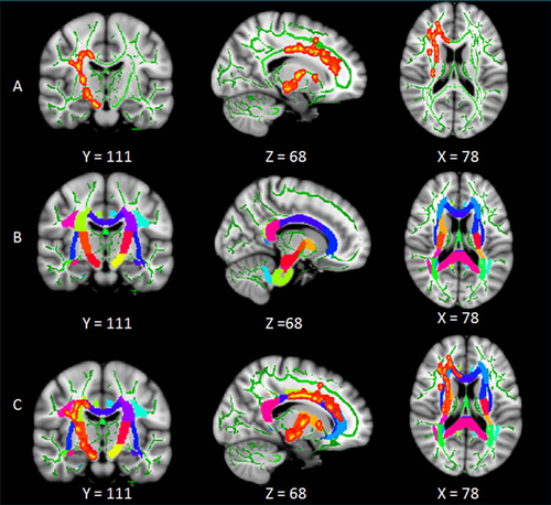
Figure 1. Tract-based analysis for whole WM tracts. A, Significant differences in WM detected from analysis of tract-based spatial statistics. Results are shown overlaid on the Montreal Neurologic Institute 152-T1 template and the mean FA skeleton (green). B, The Johns Hopkins University International Consortium of Brian Mapping Diffusion Tensor Imaging 81 atlas that covers the entire skeletons of major WM tracts is shown overlaid on the Montreal Neurologic Institute 152-T1 template. C, The overlaid region of significant clusters obtained from tract-based spatial statistics analysis and the Johns Hopkins University International Consortium of Brian Mapping Diffusion Tensor Imaging 81 atlas. The overlapping WM labeling from the Johns Hopkins University International Consortium of Brian Mapping Diffusion Tensor Imaging 81 atlas was used to extract the whole WM and to calculate the mean FA value that represents the integrity of the whole tract.
High-res (TIF) version
(Right-click and Save As)
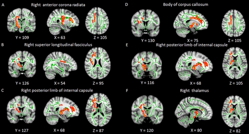
Figure 2. Significant WM clusters obtained from tract-based spatial statistics between primary insomnia and HC groups. Green represents the mean FA skeleton and red-yellow shows clusters with a significant FA reduction in the primary insomnia group (P ˂ .05, family-wise error correction). Underlying gray-scale images are the Montreal Neurologic Institute 152-T1 template. Each panel is the best axial, coronal, and sagittal view that shows the specific WM tract.
High-res (TIF) version
(Right-click and Save As)
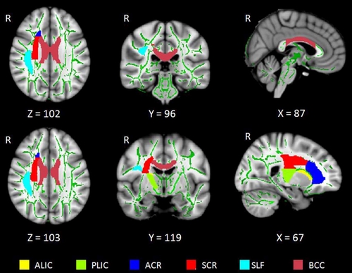
Figure 3. Distribution of the six whole WM tracts in the brain. ALIC = anterior limb of the internal capsule, ACR = anterior corona radiata, BCC = body of the corpus callosum, PLIC = posterior limb of the internal capsule, R = right side of the brain, SCR = superior corona radiata, SLF = superior longitudinal fasciculus.
High-res (TIF) version
(Right-click and Save As)
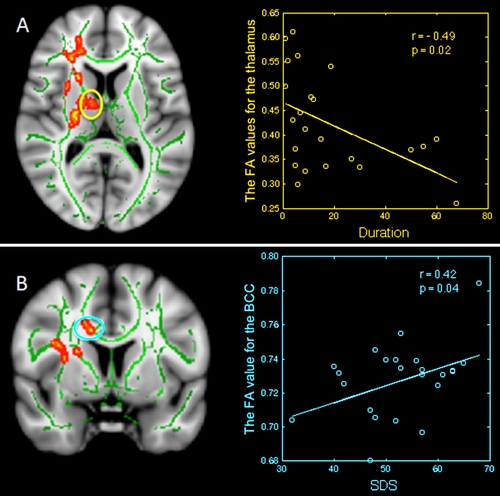
Figure 4. Relationship between mean FA and disease duration and clinical features in primary insom¬nia; region-of-interest maps (left side) are displayed with the corresponding graphs (right side). A, Sig¬nificant negative correlation between FA values for the right thalamus with disease duration in primary insomnia. B, Significant positive correlation between FA values for the body of the corpus callosum with self-rating depression scale scores (SDS) in primary insomnia. BCC = body of the corpus callosum.
High-res (TIF) version
(Right-click and Save As)