RSNA Press Release
- The goal of facial transplant surgery is to restore facial form and function in patients with devastating injuries to the face.
- Combining multiple imaging exams with computer modeling may improve surgical planning and aid in monitoring outcomes of human face transplantation.
- 3-D modeling offers critical insight into structural relationships among different facial tissues, such as skin, muscle, blood vessels and bone.
Integrated 3-D Imaging Facilitates Human Face Transplantation
Released: November 28, 2011
| Media Contacts: | RSNA Newsroom | 1-312-949-3233 |
| Before 11/26/2011 or after 12/01/2011: | RSNA Media Relations: | 1-630- 590-7762 |
| |
Linda Brooks 1-630-590-7738 lbrooks@rsna.org |
Maureen Morley 1-630-590-7754 mmorley@rsna.org |
CHICAGO—By combining conventional medical imaging with some of the same 3-D modeling techniques used in Hollywood blockbusters, researchers are offering new hope to victims of serious facial injuries. Results of a new study on human face transplantation, led by Darren M. Smith, M.D., plastic surgery resident at the University of Pittsburgh Medical Center (UPMC), were presented today at the annual meeting of the Radiological Society of North America (RSNA).
Devastating injuries or defects of the face are extremely challenging, if not impossible, to satisfactorily reconstruct by traditional surgical techniques. In face transplantation, facial tissue from a donor is transferred to reconstruct the defect, restore essential life-sustaining functions—such as breathing, chewing and speaking—and, above all, reestablish normal human appearance.
"This surgery is for patients with devastating injuries to the face, who have lost their ability to smell, eat and engage socially and have no other conventional treatment options," said Vijay S. Gorantla, M.D., Ph.D., administrative medical director of the Reconstructive Transplantation Program at UPMC.
Clearly defining and understanding the complex tissue deficits and defects that accompany devastating facial injuries like electric burns, blast wounds and accidental trauma are critical for both technical success and objective analysis of the return of function after face transplantation.
Medical imaging plays a major role in the entire spectrum of face transplantation, ranging from patient selection, donor and recipient surgical planning, and postoperative assessment of returning motor and sensory function. Face transplantation is a lengthy, complicated procedure that involves reconstruction of multiple tissues—such as skin, muscle, blood vessels, nerves and bone—by a team of surgeons.
Currently, to prepare for facial transplantation, plastic or plaster models are first created based on 3-D CT or angiographic images or reconstruction. Following this, mock cadaveric dissections are performed to allow surgeons to plan for the donor and recipient surgeries. MRI and other imaging exams may also be used to provide supplemental information.
By combining information from multiple imaging exams and creating a sophisticated 3-D computer model, the researchers for this study were better able to assess the facial structure and contours, the underlying bone, muscles, nerves and vessels, as well as the extent of damage.
Using sophisticated computer modeling software, Drs. Smith and Gorantla, along with Joseph Losee, M.D., integrated information from 3-D CT, CT angiography, MRI and high-definition tractography to create a 3-D model of the patient's head and neck anatomy. The same type of modeling technology is often used in movies to animate computer-generated characters with detailed three-dimensional human features and realistic expressions.
"We have integrated data from multiple imaging sources into a single 3-D representation that allows for real-time user interaction and modification," Dr. Smith said. "In assessing eligibility for this procedure, it is critical to understand whether the patient has enough blood vessels and bone structure to support new facial tissue. This 3-D modeling helps us customize the procedure to the patient's individual anatomy so that the donor tissue will fit like a puzzle piece onto the patient's face."
Using computer modeling, the team also overlaid the patient model with a polygon mesh of a generic human face and then customized it to the recipient facial anatomy. Dr. Smith said the ability to manipulate this 3-D facial envelope over the residual face model allows the entire surgical team to participate in planning exactly where bone, blood vessel and nerves will be cut and connected, as well as to evaluate the outcomes of reconstructive transplantation, including nerve regeneration within the transplanted facial tissue.
"The goal of face transplantation is not just structural," Dr. Gorantla said. "It is about restoring function, so that patients are once again able to chew their food, smile and regain the most important aspect of a normal face — to look human."
# # #
Note: Copies of RSNA 2011 news releases and electronic images will be available online at RSNA.org/press11 beginning Monday, Nov. 28.
RSNA is an association of more than 48,000 radiologists, radiation oncologists, medical physicists and related scientists committed to excellence in patient care through education and research. The Society is based in Oak Brook, Ill. (RSNA.org)
Editor's note: The data in these releases may differ from those in the printed abstract and those actually presented at the meeting, as researchers continue to update their data right up until the meeting. To ensure you are using the most up-to-date information, please call the RSNA Newsroom at 1-312-949-3233.
For patient-friendly information on CT and MRI, visit RadiologyInfo.org.
| Abstract: |
Video clips
- Video clip (433 KB)
Dr. Vijay Gorantla discusses the importance of new and advanced imaging technologies. - Video clip (985 KB)
Dr. Vijay Gorantla discusses the importance of combining imaging modalities. - Video clip (901 KB)
Dr. Vijay Gorantla discusses the importance of combining imaging modalities. - Video clip (441 KB)
Dr. Vijay Gorantla reveals the purpose of the study. - Video clip (481 KB)
Dr. Vijay Gorantla explains face transplantation. - Video clip (402 KB)
Dr. Vijay Gorantla explains how the face functions after transplantation. - Video clip (437 KB)
Dr. Vijay Gorantla shares the significance of the study. - Video clip (608 KB)
Dr. Vijay Gorantla shares the importance of the study. - Video clip (678 KB)
Dr. Vijay Gorantla reveals the findings of the study. - Video clip (577 KB)
Dr. Vijay Gorantla shares the next steps for the study. - Video clip (840 KB)
Dr. Vijay Gorantla compares current techniques for face transplantation with the techniques used for the study. - Video clip (429 KB)
Dr. Joseph Losee discusses the field of face transplantation. - Video clip (829 KB)
Dr. Joseph Losee describes the types of reconstructive transplantation. - Video clip (636 KB)
Dr. Joseph Losee shares the history of face transplantation. - Video clip (939 KB)
Dr. Joseph Losee explains the challenges associated with face transplantation. - Video clip (662 KB)
Dr. Joseph Losee describes how the face functions after transplantation. - Video clip (859 KB)
Dr. Joseph Losee describes how the nerves in the face function after transplantation. - Video clip (426 KB)
Dr. Darren Smith discusses the various types of imaging modalities. - Video clip (346 KB)
Dr. Darren Smith explains how models of the face are generated. - Video clip (504 KB)
Dr. Darren Smith shares the methodology of the study. - Video clip (447 KB)
Dr. Darren Smith shares the results of the study. - Video clip (742 KB)
Dr. Darren Smith reveals the findings of the study. - Video clip (507 KB)
Dr. Darren Smith discusses how facial features are reconstructed. - Video clip (543 KB)
Dr. Darren Smith shares the significant findings of the study. - Video clip (244 KB)
Laser guide mapping patient's face. - Video clip (157 KB)
Laser guide mapping patient's face. - Video clip (99 KB)
Laser machine - Video clip (863 KB)
3-D facial image
Images (.JPG format)

Figure 1: High-res (TIF) version (Right-click and Save As) |
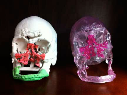
Figure 2: High-res (TIF) version (Right-click and Save As) |
 PDF
PDF