Aerobic Exercise Preserves Brain Volume and Improves Cognitive Function
Released: November 30, 2016
At A Glance
- Aerobic exercise may increase brain volume and cognitive function in people with mild cognitive impairment.
- MRI results showed increased local gray matter volume in patients who engaged in either aerobic or stretching exercises for six months, but some white matter atrophy in the stretching group.
- Cognitive testing revealed the aerobic exercise group also showed improvement in executive function after six months.
- RSNA Media Relations
1-630-590-7762
media@rsna.org - Maureen Morley
1-630-590-7754
mmorley@rsna.org - Linda Brooks
1-630-590-7738
lbrooks@rsna.org
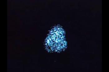
CHICAGO — Using a new MRI technique, researchers found that adults with mild cognitive impairment (MCI) who exercised four times a week over a six-month period experienced an increase in brain volume in specific, or local, areas of the brain, but adults who participated in aerobic exercise experienced greater gains than those who just stretched. The study will be presented today at the annual meeting of the Radiological Society of North America (RSNA).
"Even over a short period of time, we saw aerobic exercise lead to a remarkable change in the brain," said the study's lead investigator, Laura D. Baker, Ph.D., from Wake Forest School of Medicine (WFSM) in Winston-Salem, N.C.
The study included 35 adults with MCI participating in a randomized, controlled trial of exercise intervention. Individuals with MCI are at risk of developing Alzheimer's disease (AD), the most common form of dementia, which affects more than 5 million Americans today.
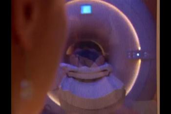
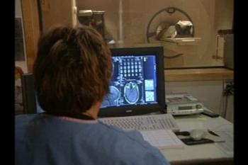
The participants were divided into two groups. Sixteen adults (average age 63 years) engaged in aerobic activity, including treadmill, stationary bike or elliptical training, four times per week for six months. A control group of 19 adults (average age 67 years) participated in stretching exercises with the same frequency. High-resolution brain MR images were acquired from all participants before and after the six-month activity period. The MRI results were compared using conventional and biomechanical metrics to measure the change in both brain volume and shape.
"We used high-resolution MR images to measure anatomical changes within areas of the brain to obtain both volumetric data and directional information," said Jeongchul Kim, Ph.D., co-investigator on the study from WFSM.
The analysis revealed that for both the aerobic and stretching groups, brain volume increased in most gray matter regions, including the temporal lobe, which supports short-term memory.
"Compared to the stretching group, the aerobic activity group had greater preservation of total brain volume, increased local gray matter volume and increased directional stretch of brain tissue," Dr. Kim said.
Among participants of the stretching group, the analysis revealed a local contraction, or atrophy, within the white matter connecting fibers. According to Dr. Kim, such directional deformation, or shape change, is partially related to volume loss, but not always.
"Directional changes in the brain without local volume changes could be a novel biomarker for neurological disease," he said. "It may be a more sensitive marker for the tiny changes that occur in a specific brain region before volumetric changes are detectable on MRI."
He said both MRI measures are important to the treatment of MCI and AD, which require the careful tracking of changes in the brain while patients engage in interventions including diet and exercise to slow the progression of the disease.
Study participants were tested to determine the effect of exercise intervention on cognitive performance. Participants in the aerobic exercise group showed statistically significant improvement in executive function after six months, whereas the stretching group did not improve.
"Any type of exercise can be beneficial," Dr. Kim said. "If possible, aerobic activity may create potential benefits for higher cognitive functioning."
Other co-authors on the study are Suzanne Craft, Ph.D., Youngkyoo Jung, Ph.D., and Christopher T. Whitlow, M.D., Ph.D.
Note: Copies of RSNA 2016 news releases and electronic images will be available online at RSNA.org/press16 beginning Monday, Nov. 28.
RSNA is an association of more than 54,000 radiologists, radiation oncologists, medical physicists and related scientists, promoting excellence in patient care and health care delivery through education, research and technologic innovation. The Society is based in Oak Brook, Ill. (RSNA.org)
Editor's note: The data in these releases may differ from those in the published abstract and those actually presented at the meeting, as researchers continue to update their data right up until the meeting. To ensure you are using the most up-to-date information, please call the RSNA Newsroom at 1-312-791-6610.
For patient-friendly information on brain MRI, visit RadiologyInfo.org.


