Obesity in Adolescence May Cause Permanent Bone Loss
Released: November 21, 2016
At A Glance
- Having a high amount of visceral fat and a low amount of muscle mass may lead to bone loss in obese adolescents.
- Bone loss during adolescence increases fracture risk later in life.
- More than one-third of children and adolescents in the U.S. are overweight or obese.
- RSNA Media Relations
1-630-590-7762
media@rsna.org - Maureen Morley
1-630-590-7754
mmorley@rsna.org - Linda Brooks
1-630-590-7738
lbrooks@rsna.org
Chicago — Teenagers who are obese may be doing irreparable damage to their bones, according to a new study being presented next week at the annual meeting of the Radiological Society of North America (RSNA).
Obesity in childhood and adolescence is associated with a number of health risks, including cardiovascular disease and diabetes. For the new study, researchers are looking at how excess weight may affect bone structure.

"While obesity was previously believed to be protective of bone health, recent studies have shown a higher incidence of forearm fractures in obese youths," said the study's lead author, Miriam A. Bredella, M.D., radiologist at Massachusetts General Hospital and associate professor of radiology at Harvard Medical School in Boston.
Dr. Bredella and colleagues set out to determine the relationship between adolescent obesity and bone structure. The researchers have recruited 23 obese adolescents with a mean age of 17 years and a mean body mass index (BMI) of 44 kg/m2 for the ongoing study.
"Adolescence is the time where we accrue our peak bone mass, so bone loss during this time is a serious problem," Dr. Bredella said. "We know from other chronic states that lead to bone loss in adolescence, such as anorexia nervosa, that increased fracture risk persists in adulthood, even after normalization of body weight. Therefore, it is important to address this problem early on."
The researchers performed 3D HR-pQCT—a type of computed tomography exam designed specifically for measuring bone mineral density and bone microarchitecture in the arms and legs—to determine the bone structure of the distal radius, an area of the forearm near the wrist. They also performed dual-energy x-ray absorptiometry (DXA) exams to determine body composition, including lean mass and visceral fat mass. Visceral fat is the deep fat in the abdomen that surrounds the internal organs.
"There are several mechanisms by which visceral fat exerts negative effects on bone," Dr. Bredella said. "Visceral fat secretes substances that promote chronic inflammation, and chronic inflammation stimulates formation of osteoclasts, which are the cells that resorb or break-down bone. In addition, vitamin D, which is important for bone health, is soluble in adipose tissue and gets trapped within fat cells."
She noted that growth hormone, which is important for bone health, is also lower in adolescents with visceral obesity.
The study results showed that BMI was positively associated with cortical thickness and area. Cortical bone is dense and compact and forms the outer shell of most bones. Visceral fat mass was positively associated with cortical porosity.
Lean mass was positively associated with trabecular density, volume and integrity. Trabecular bone is a spongy inner layer of bone that provides support and flexibility.
The findings suggest that having a high amount of visceral fat coupled with a low amount of muscle mass puts adolescents at risk for weakened bone structure.
"The best way to prevent bone loss is a healthy diet that contains adequate amounts of calcium and vitamin D, along with sufficient exercise, as we have shown in our study that muscle mass is good for bone health," Dr. Bredella said.
According to the Centers for Disease Control and Prevention (CDC), obesity has more than quadrupled in adolescents over the past 30 years. It is estimated that more than one-third of children and adolescents in the U.S. are overweight or obese.
Co-authors on the study are Fatima C. Stanford, M.D., M.P.H., M.P.A., Vibha Singhal, M.D., M.B.B.S., Stijn A. Bos, B.S., Ryan Woolley, B.S., Alexander Toth, B.S., and Madhusmita Misra, M.D., M.P.H.
Note: Copies of RSNA 2016 news releases and electronic images will be available online at RSNA.org/press16 beginning Monday, Nov. 28.
RSNA is an association of more than 54,000 radiologists, radiation oncologists, medical physicists and related scientists, promoting excellence in patient care and health care delivery through education, research and technologic innovation. The Society is based in Oak Brook, Ill. (RSNA.org)
Editor's note: The data in these releases may differ from those in the published abstract and those actually presented at the meeting, as researchers continue to update their data right up until the meeting. To ensure you are using the most up-to-date information, please call the RSNA Newsroom at 1-312-791-6610.
For patient-friendly information on CT and DXA, visit RadiologyInfo.org.
Images (.JPG and .TIF format)
and Video clips (.mp4 format)
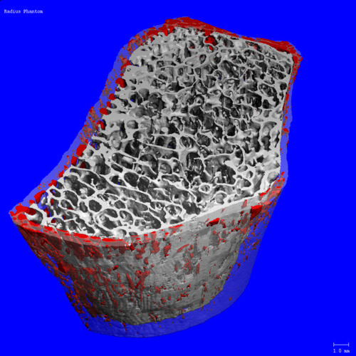
Figure 1. Image acquired using a SCANCO Medical XtremeCT scanner.
High-res (TIF) version
(Right-click and Save As)
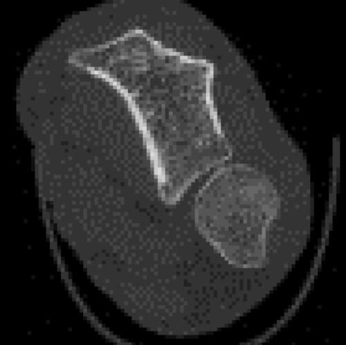
Figure 2. Clinical measurement of an arm cross-section acquired with a SCANCO Medical XtremeCT scanner.
High-res (TIF) version
(Right-click and Save As)
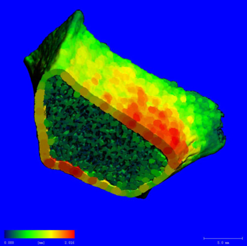
Figure 3. Thickness map of the radius bone (the smaller of the two bones of the forearm) acquired with a SCANCO Medical XtremeCT scanner.
High-res (TIF) version
(Right-click and Save As)

Figure 4. Image acquired using a SCANCO Medical XtremeCT scanner.
High-res (TIF) version
(Right-click and Save As)
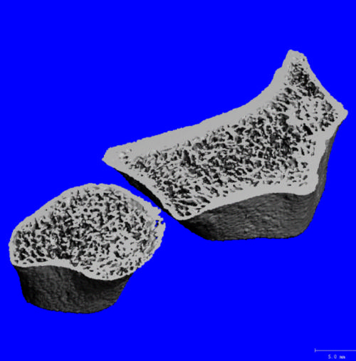
Figure 5. Clinical measurement of the radius and ulna (the two bones of the forearm) segmented (separated) acquired with a SCANCO Medical XtremeCT scanner).
High-res (TIF) version
(Right-click and Save As)

Figure 6. Photograph of a SCANCO Medical XtremeCT scanner.
High-res (TIF) version
(Right-click and Save As)
Video 1. Dr. Bredella discusses what kind of fat is most damaging to bone strength.
Download.mp4
(Right-click and Save As)
Video 2. Dr. Bredella discusses how to prevent bone degeneration.
Download.mp4
(Right-click and Save As)
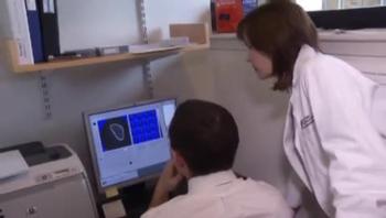
Video 3. Video clip showing Dr. Bredella consulting with another radiologist in a reading room.
Download.mp4
(Right-click and Save As)
Video 4. Video clip showing a radiologic technologist putting a patient’s arm in a computed tomography (CT) forearm scanner.
Download.mp4
(Right-click and Save As)
Video 5. Video clip showing Dr. Bredella consulting with a radiologic technologist.
Download.mp4
(Right-click and Save As)
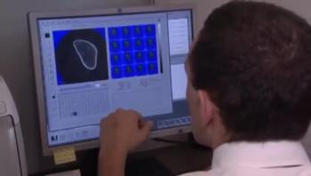
Video 6. Video clip showing a radiologic technologist reviewing images.
Download.mp4
(Right-click and Save As)
Video 8. Miriam Bredella, M.D., describes the findings of her study
Download.mp4
(Right-click and Save As)
Video 9. Miriam Bredella, M.D., discusses the effect adolescent obesity has on bones.
Download.mp4
(Right-click and Save As)
Video 10. Miriam Bredella, M.D., explains why the study is important.
Download.mp4
(Right-click and Save As)