RSNA Press Release
- Mammography screening using a technique called “photon counting” offers high diagnostic performance.
- Researchers compared the screening performance of a direct radiography photon-counting scan system with those of statewide operating screening units using different digital technologies.
- The higher cancer detection resulting from the use of the photon-counting scan system is due to high detection of both small, invasive cancers and ductal carcinoma in situ.
Novel Technique Increases Detection Rate in Screening Mammography
Released: February 4, 2014
| Media Contacts: | |
| RSNA Media Relations: | 1-630-590-7762 |
| Linda Brooks 1-630-590-7738 lbrooks@rsna.org |
Maureen Morley 1-630-590-7754 mmorley@rsna.org |


OAK BROOK, Ill. — Digital mammography screening with new photon-counting technique offers high diagnostic performance, according to a study published online in the journal Radiology.
As mammography screening has shifted to digital technology, a range of computed radiography (CR) and direct radiography (DR) systems have emerged. The photon-counting technique is a promising DR approach that uses a unique detector to decrease scattered radiation and noise, enabling dose reduction and making it a promising tool for screening.
"In population-based mammography screening, dose reducing techniques that don't compromise outcome parameters are desirable," said Walter Heindel, M.D., from the Department of Clinical Radiology at the University Hospital Muenster in Muenster, Germany.
For the study, Dr. Heindel and colleagues analyzed data from the mammography screening program in North Rhine-Westphalia, the most populous state in Germany. They compared the screening performance of a DR photon-counting scan system with those of statewide operating screening units using different digital technologies. During the study period (2009 – 2010), 13,312 women were examined using the photon-counting system, and 993,822 women were screened with either CR or DR systems alone.
The DR photon-counting scan system had a cancer detection rate of 0.76 percent for subsequent screening, compared with 0.59 percent for the other screening units. The recall rate was 5.4 percent for the photon-counting method and 3.4 percent for the other methods.
"The higher cancer detection resulting from the use of the DR photon-counting scan system is due to high detection of both small, invasive cancers and ductal carcinoma in situ," Dr. Heindel said.
The photon-counting technique had almost twice the detection rate of other methods for ductal carcinoma in situ (DCIS), an early, noninvasive form of disease. It had a higher DCIS detection rate than the statewide units and the conventional DR subgroup.
In addition, the mean average glandular radiation dose of the DR photon-counting scan system was significantly lower than the conventional DR systems with the individually used parameters of the automatic exposure control.
The study's large size distinguishes it from previous studies that compared the DR photon-counting scan system's performance with other approaches.
"To our knowledge, the study is different from previous ones as we examined the performance of the DR photon-counting scan mammography on a larger database with consideration of multiple parameters of screening," said study coauthor Stefanie Weigel, M.D., from the University Hospital Muenster.
The photon-counting technique also offers lateral dose modulation during the image acquisition, which can help account for differences in breast density. Cancer often is more difficult to detect in women with dense breasts.
"The innovative photon-counting technique offers further research potential," Dr. Heindel said. "One future research direction is the application of spectral imaging for quantification of breast glandular tissue, addressing the problem of breast density."
# # #
"Digital Mammography Screening with Photon-counting Technique: Can a High Diagnostic Performance Be Realized at Low Mean Glandular Dose?" Collaborating with Drs. Heindel and Weigel were Shoma Berkemeyer, Ph.D., Ralf Girnus, M.Sc., Alexander Sommer, M.Sc., and Horst Lenzen, M.Sc.
Radiology is edited by Herbert Y. Kressel, M.D., Harvard Medical School, Boston, Mass., and owned and published by the Radiological Society of North America, Inc. (http://radiology.rsna.org/)
RSNA is an association of more than 53,000 radiologists, radiation oncologists, medical physicists and related scientists, promoting excellence in patient care and health care delivery through education, research and technologic innovation. The Society is based in Oak Brook, Ill. (RSNA.org)
For patient-friendly information on digital mammography, visit RadiologyInfo.org.
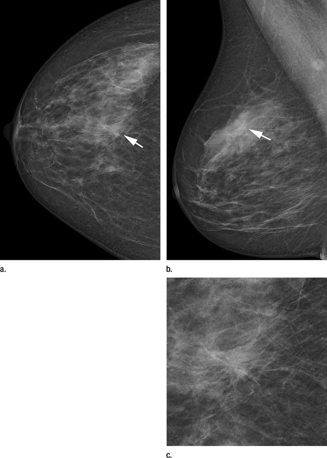
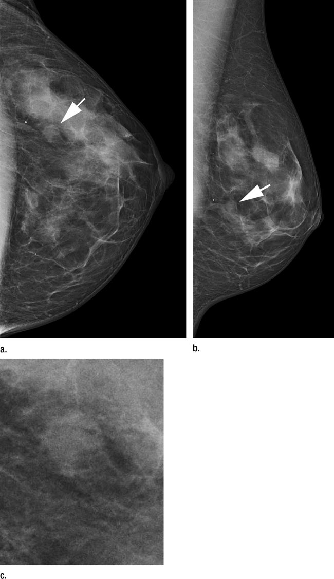
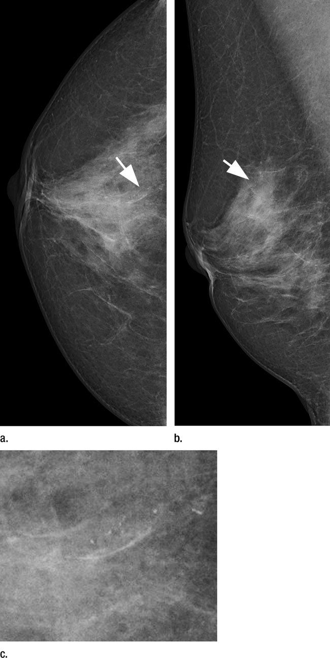
Images
|
High-res (TIF) version (Right-click and Save As) |
High-res (TIF) version (Right-click and Save As) |
High-res (TIF) version (Right-click and Save As) |
 PDF
PDF