RSNA Press Release
- Ultrasound with elastography may improve skin cancer detection.
- Elastography identifies cancer based on skin elasticity and can assess the extent and depth of the skin lesion.
- More than one million cases of skin cancer are diagnosed in the U.S. each year.
Special Ultrasound Accurately Identifies Skin Cancer
Released: December 1, 2009
| Media Contacts: | RSNA Newsroom | 1-312-949-3233 |
| Before 11/28/09 or after 12/03/09: | RSNA Media Relations: | 1-630- 590-7762 |
| |
Linda Brooks 1-630-590-7738 lbrooks@rsna.org |
Maureen Morley 1-630-590-7754 mmorley@rsna.org |
CHICAGO — High-frequency ultrasound with elastography can help differentiate between cancerous and benign skin conditions, according to a study presented today at the annual meeting of the Radiological Society of North America (RSNA).
"High-frequency ultrasound with elastography has the potential to improve the efficiency of skin cancer diagnosis," said lead author Eliot L. Siegel, M.D., vice chairman of the Department of Radiology at the University of Maryland School of Medicine (UMSM) in Baltimore. "It successfully delineated the extent of lesions and was able to provide measurable differentiation among a variety of benign and malignant lesions."
There are more than one million cases of skin cancer diagnosed in the U.S. every year, according to the American Cancer Society. Melanoma, the most serious type of skin cancer, will account for about 68,720 cases of skin cancer and 11,590 deaths in 2009, despite the fact that with early detection it is highly curable.
Suspicious skin lesions are typically diagnosed by dermatologists and biopsied based on their surface appearance and characteristics. Unfortunately, even to experienced dermatologists, benign and malignant lesions often appear similar visually and on physical examination, and some malignant lesions may have a benign appearance, especially in their early stages. It is not uncommon for patients to have one or more lesions that appear concerning.
"Dermatologists tend to biopsy any lesions that seem visually suspicious for disease," said coauthor Bahar Dasgeb, M.D., from the Department of Dermatology at Wayne State University in Detroit and Pinkus Dermatopathology Lab in Monroe, Michigan. "Consequently, many benign lesions are needlessly biopsied in order to avoid the risk of missing a potentially deadly melanoma."
Elastography was found to distinguish between benign and malignant lesions not by their visible appearance but by measuring their elasticity or stiffness. Since malignancies are stiffer than benign growths, elastography, when added to high-frequency ultrasound imaging of the skin, has potential to improve the accuracy of traditional clinical diagnosis of skin cancers and, in some cases, eliminate unnecessary biopsies of benign skin lesions. The procedure is noninvasive, convenient and inexpensive.
For the study, researchers used an ultra high-frequency ultrasound system to image 40 patients with a variety of malignant and nonmalignant, or benign, skin lesions. Malignant tumors included squamous cell carcinoma, basal cell carcinoma and melanoma. Benign lesions included dermatofibroma, a noncancerous growth containing scar tissue, and lipoma, a noncancerous tumor composed of fatty tissue.
The researchers calculated the ratio of elasticity between normal skin and the adjacent skin lesion, and used laboratory analysis to confirm their diagnoses. Cystic lesions, which are not malignant, demonstrated high levels of elasticity, while malignant lesions were significantly less elastic. The elasticity ratio of normal skin to the various skin lesions ranged from 0.04 to 0.3 for cystic skin lesions to above 10.0 for malignant lesions.
In addition, high-frequency ultrasound with elastography allows for accurate characterization of the extent and depth of the lesion below the surface, which can aid physicians in treatment.
"The visualized portion of a skin lesion can be just the tip of the iceberg, and most dermatologists operate 'blindly' beyond what they can see on the surface," Dr. Siegel said. "High-frequency ultrasound provides almost microscopic resolution and enables us to get size, shape and extent of the lesion prior to biopsy."
# # #
Note: Copies of RSNA 2009 news releases and electronic images will be available online at RSNA.org/press09 beginning Monday, Nov. 30.
RSNA is an association of more than 44,000 radiologists, radiation oncologists, medical physicists and related scientists committed to excellence in patient care through education and research. The Society is based in Oak Brook, Ill. (RSNA.org)
Editor's note: The data in these releases may differ from those in the printed abstract and those actually presented at the meeting, as researchers continue to update their data right up until the meeting. To ensure you are using the most up-to-date information, please call the RSNA Newsroom at 1-312-949-3233.
For patient-friendly information on ultrasound, visit RadiologyInfo.org.
| Abstract: |
Images (.JPG format)
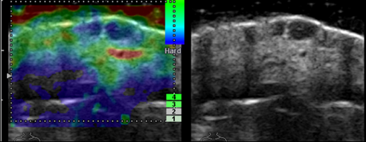 Figure 1. An elastogram (left) and ultrasound image (right) showing squamous cell carcinoma of the skin. High-res (TIF) version (Right-click and Save As) |
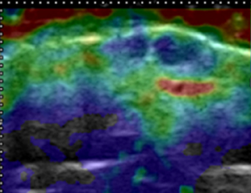 Figure 2. An elastogram showing squamous cell carcinoma of the skin. High-res (TIF) version (Right-click and Save As) |
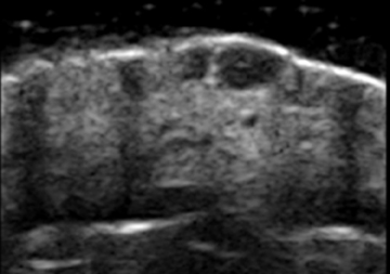 Figure 3. An ultrasound image showing squamous cell carcinoma of the skin. High-res (TIF) version (Right-click and Save As) |
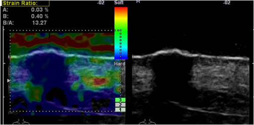 Figure 4. An elastogram (left) and ultrasound (right) showing squamous cell carcinoma of the skin. High-res (TIF) version (Right-click and Save As) |
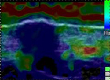 Figure 5. An elastogram showing squamous cell carcinoma of the skin. High-res (TIF) version (Right-click and Save As) |
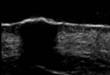 Figure 6. An ultrasound image showing squamous cell carcinoma of the skin. High-res (TIF) version (Right-click and Save As) |
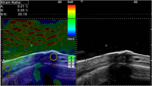 Figure 7. An elastogram (left) and ultrasound image (right) showing squamous cell carcinoma of the skin. High-res (TIF) version (Right-click and Save As) |
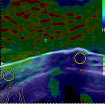 Figure 8. An elastogram showing squamous cell carcinoma of the skin. High-res (TIF) version (Right-click and Save As) |
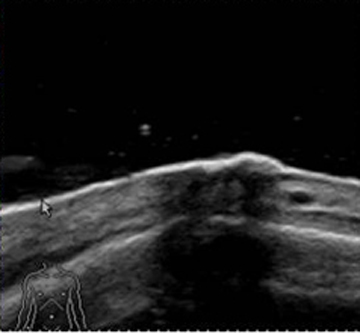 Figure 9. An ultrasound image showing squamous cell carcinoma of the skin. High-res (TIF) version (Right-click and Save As) |
 Figure 10. An elastogram (left) and ultrasound image (right) showing skin cancer. High-res (TIF) version (Right-click and Save As) |
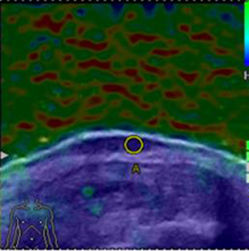 Figure 11. An elastogram showing skin cancer. High-res (TIF) version (Right-click and Save As) |
 Figure 12. An ultrasound image showing skin cancer. High-res (TIF) version (Right-click and Save As) |
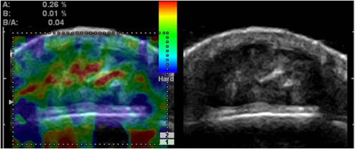 Figure 13. An elastogram (left) and ultrasound image (right) showing a benign cystic proliferation. High-res (TIF) version (Right-click and Save As) |
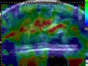 Figure 14. An elastogram showing a benign cystic proliferation. High-res (TIF) version (Right-click and Save As) |
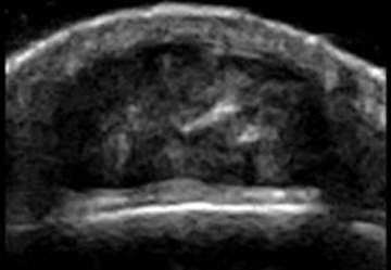 Figure 15. An ultrasound image showing a benign cystic proliferation. High-res (TIF) version (Right-click and Save As) |
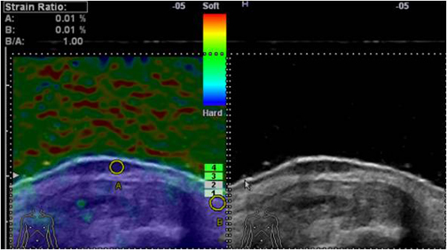 Figure 16. An elastogram (left) and ultrasound image (right) showing a premalignant lesion. High-res (TIF) version (Right-click and Save As) |
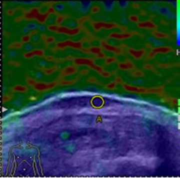 Figure 17. An elastogram showing a premalignant lesion. High-res (TIF) version (Right-click and Save As) |
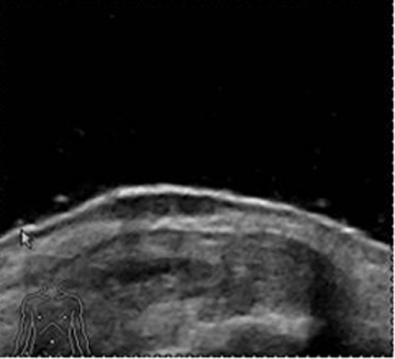 Figure 18. An ultrasound image showing a premalignant lesion. High-res (TIF) version (Right-click and Save As) |
 PDF
PDF