RSNA Press Release
- Radiologists have begun using a minimally invasive, image-guided technique to detect and remove objects inserted by teens into their arms, hands, feet, ankles and necks.
- This is the first study to report on an emerging condition known as self-embedding disorder.
- Self-embedded objects removed included needles, staples, paper clips, wood, stone, glass, pencil lead and a crayon.
- Self-injury has been reported in 13 to 24 percent of high school students in the U.S. and Canada.
Radiologists Diagnose and Treat Self-Embedding Disorder in Teens
Released: December 3, 2008
| Media Contacts: | RSNA Newsroom | 1-312-949-3233 |
| Before 11/29/08 or after 12/04/08: | RSNA Media Relations: | (630) 590-7762 |
| |
Maureen Morley 1-630-590-7754 mmorley@rsna.org |
Linda Brooks 1-630-590-7738 lbrooks@rsna.org |
CHICAGO — Minimally invasive, image-guided treatment is a safe and precise method for removal of self-inflicted foreign objects from the body, according to the first report on "self-embedding disorder," or self-injury and self-inflicted foreign body insertion in adolescents. The findings will be presented today at the annual meeting of the Radiological Society of North America (RSNA).
"Radiologists are in a unique position to be the first to detect self-embedding disorder, make the appropriate diagnosis and mobilize the healthcare system for early and effective intervention and treatment," said the study's principal investigator, William E. Shiels II, D.O., chief of the Department of Radiology at Nationwide Children’s Hospital in Columbus, Ohio.
Self-injury, or self-harm, refers to a variety of behaviors in which a person intentionally inflicts harm to his or her body without suicidal intent. It is a disturbing trend among U.S. adolescents, particularly girls. Prevalence is unknown because many cases go unreported, but recent studies have reported that 13 to 24 percent of high school students in the U.S. and Canada have practiced deliberate self-injury at least once. More common forms of self-injury include cutting of the skin, burning, bruising, hair pulling, breaking bones or swallowing toxic substances. In cases of self-embedding disorder, objects are used to puncture the skin or are embedded into the wound after cutting.
Dr. Shiels and colleagues studied 19 episodes of self-embedding injury in 10 adolescent girls, age 15 to 18. Using ultrasound and/or fluoroscopic guidance, interventional pediatric radiologists removed 52 embedded foreign objects from nine of the patients. The embedded objects included metal needles, metal staples, metal paperclips, glass, wood, plastic, graphite (pencil lead), crayon and stone. The objects were embedded during injuries to the arms, ankles, feet, hands and neck. One patient had self-embedded 11 objects, including an unfolded metal paperclip more than six inches in length.
Ultrasound guidance allowed the researchers to detect the presence and location of wood, crayons and plastic objects, not detectable on x-ray examinations. Removal was performed through small incisions in the skin that left little or no scarring and was successful in all cases, without fragmentation or complications.
"This technique offers surgeons and emergency physicians a safe and effective alternative for removal of foreign bodies, including objects at risk for fragmentation during traditional operative techniques," said co-author Adam Young, B.S. "The small incision minimizes scarring and deformity, which is key for the self-esteem of this unique, high-risk group of patients."
Co-authors are James Murakami, M.D., Brian Coley, M.D., and Mark Hogan, M.D.
# # #
RSNA is an association of more than 42,000 radiologists, radiation oncologists, medical physicists and related scientists committed to excellence in patient care through education and research. The Society is based in Oak Brook, Ill. (RSNA.org)
Editor's note: The data in these releases may differ from those in the printed abstract and those actually presented at the meeting, as researchers continue to update their data right up until the meeting. To ensure you are using the most up-to-date information, please call the RSNA Newsroom at 1-312-949-3233.
For patient-friendly information on interventional radiology procedures, visit RadiologyInfo.org.
| Abstract: |
Images (.JPG format)
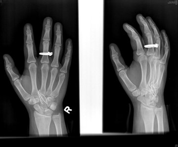 Figure 1. This x-ray image illustrates 3 metal staples embedded in the hand of a teenage girl High-res (TIF) version (Right-click and Save As) |
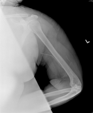 Figure 2. This x-ray image illustrates 8 metal pieces embedded in the left arm of a teenage girl High-res (TIF) version (Right-click and Save As) |
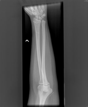 Figure 3. This x-ray image illustrates metal pieces embedded in the wrist of a teenage girl High-res (TIF) version (Right-click and Save As) |
 PDF
PDF