RSNA Press Release
- Researchers using fMRI have found that people with stress-related psychiatric disorders have difficulty suppressing traumatic memories.
- The brain’s prefrontal cortex, which controls processing of memories, is dysfunctional in patients with stress-related psychiatric disorders.
- fMRI is an important tool in understanding psychiatric disorders.
Stress-Related Disorders Affect Brain's Processing of Memory
Released: December 3, 2008
| Media Contacts: | RSNA Newsroom | 1-312-949-3233 |
| Before 11/29/08 or after 12/04/08: | RSNA Media Relations: | (630) 590-7762 |
| |
Maureen Morley 1-630-590-7754 mmorley@rsna.org |
Linda Brooks 1-630-590-7738 lbrooks@rsna.org |
CHICAGO — Researchers using functional MRI (fMRI) have determined that the circuitry in the area of the brain responsible for suppressing memory is dysfunctional in patients suffering from stress-related psychiatric disorders. Results of the study will be presented today at the annual meeting of the Radiological Society of North America (RSNA).
"For patients with major depression and other stress-related disorders, traumatic memories are a source of anxiety," said Nivedita Agarwal, M.D., radiology resident at the University of Udine in Italy, where the study is being conducted, and research fellow at the Brain Imaging Center of McLean Hospital, Department of Psychiatry at Harvard Medical School in Boston. "Because traumatic memories are not adequately suppressed by the brain, they continue to interfere with the patient’s life."
Dr. Agarwal and colleagues used brain fMRI to explore alterations in the neural circuitry that links the prefrontal cortex to the hippocampus, while study participants performed a memory task. Participants included 11 patients with major depression, 13 with generalized anxiety disorder, nine with panic attack disorders, five with borderline personality disorder and 21 healthy individuals. All patients reported suffering varying degrees of stressful traumatic events, such as sexual or physical abuse, difficult relationships or "mobbing" — a type of bullying or harassment — at some point in their lives.
After reviewing a list of neutral word pairs, each participant underwent fMRI. During imaging, they were presented with one of the words and asked to either recall or to suppress the memory of its associated word.
The fMRI images revealed that the prefrontal cortex, which controls the suppression and retrieval of memories processed by the hippocampus, showed abnormal activation in the patients with stress-related disorders compared to the healthy controls. During the memory suppression phase of the test, patients with stress-related disorders showed greater activation in the hippocampus, suggesting that insufficient activation of the prefrontal cortex could be the basis for inadequate suppression of unwanted traumatic memories stored in the hippocampus.
"These data suggest that the mechanism for memory suppression is dysfunctional in patients with stress-related disorders primarily because of an alteration of the prefrontal cortex," Dr. Agarwal said. "These patients often complain of poor memory, which might in part be attributed to this altered circuitry," she added.
According to Dr. Agarwal, fMRI is an important tool in understanding the neurobiological basis of psychiatric disorders and in identifying imaging markers to psychiatric disease, helping clinicians target specific parts of the brain for treatment.
The study's principal investigator is Paolo Brambilla, M.D., Ph.D. Co-authors are Monica Baiano, M.D., Ph.D., Massimo Bazzocchi, M.D., Giuseppe Como, M.D., and Marta Maieron, Ph.D.
# # #
RSNA is an association of more than 42,000 radiologists, radiation oncologists, medical physicists and related scientists committed to excellence in patient care through education and research. The Society is based in Oak Brook, Ill. (RSNA.org)
Editor's note: The data in these releases may differ from those in the printed abstract and those actually presented at the meeting, as researchers continue to update their data right up until the meeting. To ensure you are using the most up-to-date information, please call the RSNA Newsroom at 1-312-949-3233.
For patient-friendly information on fMRI, visit RadiologyInfo.org.
| Abstract: |
Images (.JPG format)
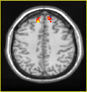 Figure 1. Brain fMRI image illustrating less activation of the prefrontal cortex (PFC) during the memory suppression phase. High-res (TIF) version (Right-click and Save As) |
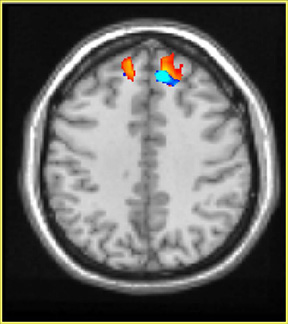 Figure 2. Brain fMRI image illustrating activation of the prefrontal cortex (PFC) during the memory retrieval phase. High-res (TIF) version (Right-click and Save As) |
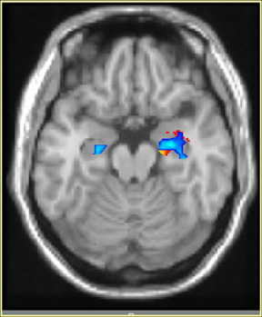 Figure 3. Brain fMRI image illustrating activation of the hippocampus during the memory suppression phase. High-res (TIF) version (Right-click and Save As) |
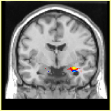 Figure 4. Brain fMRI image of a healthy patient's hippocampus during the memory retrieval phase. High-res (TIF) version (Right-click and Save As) |
 PDF
PDF