RSNA Press Release
- Breast-specific gamma imaging (BSGI) is an effective method of detecting hard-to-find breast cancer.
- BSGI identifies cancers not visible on mammograms and not found by clinical examination.
- BSGI findings are based on the metabolic activity of cancer cells.
New Breast Imaging Technology Targets Hard-to-Detect Cancers
Released: December 2, 2008
| Media Contacts: | RSNA Newsroom | 1-312-949-3233 |
| Before 11/29/08 or after 12/04/08: | RSNA Media Relations: | (630) 590-7762 |
| |
Maureen Morley 1-630-590-7754 mmorley@rsna.org |
Linda Brooks 1-630-590-7738 lbrooks@rsna.org |
CHICAGO — Breast-specific gamma imaging (BSGI) is effective in the detection of cancers not found on mammograms or by clinical exam, according to a study presented today at the annual meeting of the Radiological Society of North America (RSNA).
"BSGI can identify the most difficult to detect breast cancer—invasive lobular carcinoma," said lead author Rachel F. Brem, M.D., professor of radiology and director of the Breast Imaging and Interventional Center at The George Washington University Medical Center in Washington, D.C. "It also can help us detect additional lesions of all types of breast cancer in women whose mammograms show only one suspicious lesion."
Breast cancer affects more women than any other non-skin cancer and, according to the American Cancer Society, accounts for more than 40,000 deaths annually in the U.S. Most experts agree that the best way to decrease breast cancer mortality is through early detection using mammography and clinical breast exam. However, some cancers are difficult to detect with mammography and clinical exam, particularly in the earliest stage when treatment is most effective.
While mammography findings are characterized by the difference in appearance between normal and suspicious breast tissue, BSGI findings are based on how cancerous cells function.
"It is this physiological approach to breast cancer diagnosis that allows for improved cancer detection," Dr. Brem said.
BSGI is an emerging molecular imaging technology using a high-resolution gamma camera that allows for imaging with very mild compression of the breast along with an injection of a low-dose nuclear material called a radiotracer, which is absorbed by the cells. Because cancerous cells have a higher rate of metabolic activity, the tracer is taken up by these cells at a higher level than in normal cells.
Dr. Brem and colleagues reviewed the records of 159 women with at least one suspicious or cancerous lesion found by mammography or physical exam, who had undergone BSGI to determine if additional lesions were present.
BSGI results showed an additional suspicious lesion missed by mammography and physical exam in 46 (29 percent) of the women. In 14 (36 percent) of the 39 women who underwent biopsy, the newly discovered lesions were cancerous.
"The data suggest that BSGI allows for the diagnosis of more and earlier breast cancers," Dr. Brem said.
Dr. Brem pointed out that BSGI is not meant to replace mammography, but to be used as an adjunct to mammography. "It is an excellent tool for locating difficult-to-detect cancers and for screening high-risk women who have normal mammograms and physical examination," she said.
# # #
Disclosure: Dr. Brem is on the board of directors of iCAD, Inc., a board member of Dilon Technologies LLC and a consultant for Orbotech Ltd.
RSNA is an association of more than 42,000 radiologists, radiation oncologists, medical physicists and related scientists committed to excellence in patient care through education and research. The Society is based in Oak Brook, Ill. (RSNA.org)
Editor's note: The data in these releases may differ from those in the printed abstract and those actually presented at the meeting, as researchers continue to update their data right up until the meeting. To ensure you are using the most up-to-date information, please call the RSNA Newsroom at 1-312-949-3233.
For patient-friendly information on breast imaging, visit RadiologyInfo.org.
| Abstract: |
Images (.JPG format)
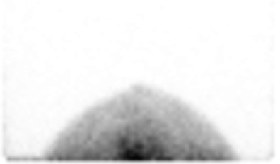 Figure 1. Image taken using breast-specific gamma imaging (BSGI). High-res (TIF) version (Right-click and Save As) |
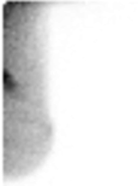 Figure 2. Image taken using breast-specific gamma imaging (BSGI). High-res (TIF) version (Right-click and Save As) |
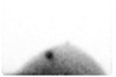 Figure 3. Image taken using breast-specific gamma imaging (BSGI). High-res (TIF) version (Right-click and Save As) |
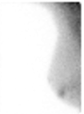 Figure 4. Image taken using breast-specific gamma imaging (BSGI). High-res (TIF) version (Right-click and Save As) |
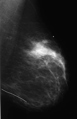 Figure 5. Image taken using breast-specific gamma imaging (BSGI). High-res (TIF) version (Right-click and Save As) |
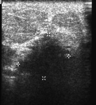 Figure 6. Image taken using breast-specific gamma imaging (BSGI). High-res (TIF) version (Right-click and Save As) |
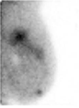 Figure 7. Image taken using breast-specific gamma imaging (BSGI). High-res (TIF) version (Right-click and Save As) |
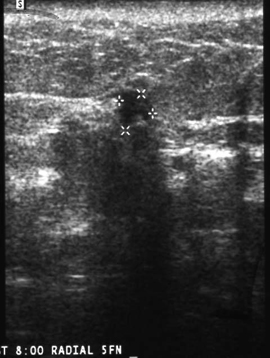 Figure 8. Image taken using breast-specific gamma imaging (BSGI). High-res (TIF) version (Right-click and Save As) |
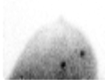 Figure 9. Image taken using breast-specific gamma imaging (BSGI). High-res (TIF) version (Right-click and Save As) |
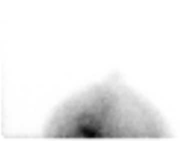 Figure 10. Image taken using breast-specific gamma imaging (BSGI). High-res (TIF) version (Right-click and Save As) |
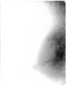 Figure 11. Image taken using breast-specific gamma imaging (BSGI). High-res (TIF) version (Right-click and Save As) |
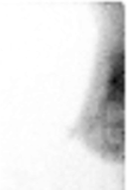 Figure 12. Image taken using breast-specific gamma imaging (BSGI). High-res (TIF) version (Right-click and Save As) |
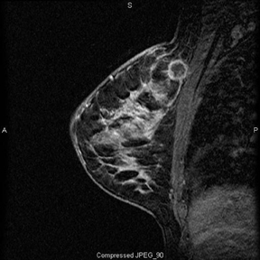 Figure 13. Image taken using breast-specific gamma imaging (BSGI). High-res (TIF) version (Right-click and Save As) |
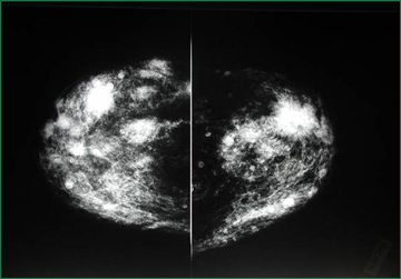 Figure 14. Image taken using breast-specific gamma imaging (BSGI). High-res (TIF) version (Right-click and Save As) |
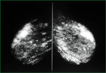 Figure 15. Image taken using breast-specific gamma imaging (BSGI). High-res (TIF) version (Right-click and Save As) |
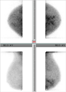 Figure 16. Image taken using breast-specific gamma imaging (BSGI). High-res (TIF) version (Right-click and Save As) |
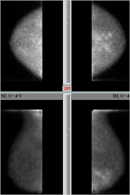 Figure 17. Image taken using breast-specific gamma imaging (BSGI). High-res (TIF) version (Right-click and Save As) |
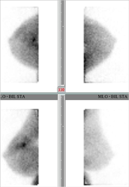 Figure 18. Image taken using breast-specific gamma imaging (BSGI). High-res (TIF) version (Right-click and Save As) |
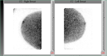 Figure 19. An example breast-specific gamma imaging (BSGI) image of a patient's left and right breasts. High-res (TIF) version (Right-click and Save As) |
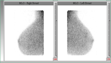 Figure 20. An example breast-specific gamma imaging (BSGI) image of a patient's left and right breasts. High-res (TIF) version (Right-click and Save As) |
 Figure 21. Gamma camera used for breast-specific gamma imaging (BSGI). High-res (TIF) version (Right-click and Save As) |
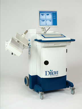 Figure 22. Gamma camera used for breast-specific gamma imaging (BSGI). High-res (TIF) version (Right-click and Save As) |
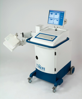 Figure 23. Gamma camera used for breast-specific gamma imaging (BSGI). High-res (TIF) version (Right-click and Save As) |
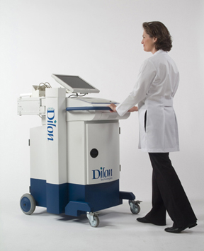 Figure 24. Gamma camera used for breast-specific gamma imaging (BSGI). High-res (TIF) version (Right-click and Save As) |
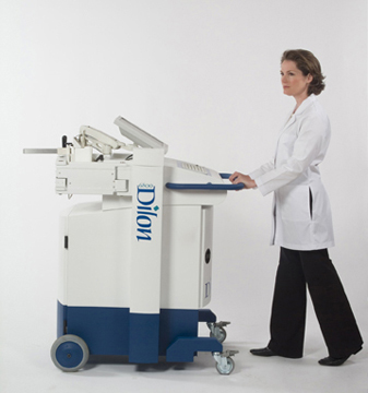 Figure 25. Gamma camera used for breast-specific gamma imaging (BSGI). High-res (TIF) version (Right-click and Save As) |
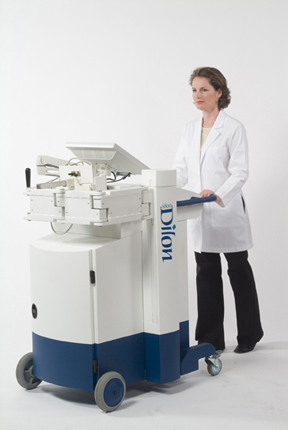 Figure 26. Gamma camera used for breast-specific gamma imaging (BSGI). High-res (TIF) version (Right-click and Save As) |
|
|
 PDF
PDF