RSNA Press Release
- The left hemisphere of the infant brain activates in response to speech, a response previously unexpected until puberty.
- This is the first time functional MRI (fMRI) has been used to study infants.
- fMRI is noninvasive and uses no ionizing radiation.
All Sides Are Not Created Equal as Babies Process Speech
Released: December 2, 2003
|
Media Contacts: |
Heather Babiar or Maureen Morley | (630) 590-7762 |
|
Heather Babiar (630) 590-7738 hbabiar@rsna.org |
Maureen Morley (630) 590-7754 mmorley@rsna.org |
CHICAGO —For the first time, researchers have used functional magnetic resonance imaging (fMRI) to investigate infant brain activity in response to speech and have found that, almost from birth, the brain's left hemisphere plays the leading role in processing most language functions. These preliminary findings, presented today at the 89th Scientific Assembly and Annual Meeting of the Radiological Society of North America (RSNA), challenge the previously held belief that left-hemisphere dominance isn't fully developed until puberty.
"Language lateralization seems to be established almost from birth," said Shantanu Sinha, Ph.D., associate professor of radiology at the David Geffen School of Medicine at University of California Los Angeles, where the study is ongoing. Lateralization is the localization of a function, such as speech, to the right or left side of the brain.
"To the best of our knowledge, this is the first time fMRI has been used to study infants," Dr. Sinha said. "Using fMRI, we can noninvasively study the neuronal response of newborns to stimulation of different kinds, without any ionizing radiation or pharmaceutical injections."
As part of a larger, longitudinal study monitoring the cognitive development of infants with brain injury from birth to age 2, the researchers performed fMRI exams on 42 infants with documented brain injury. The fMRI analysis only included cases where both sides of the brain were equally capable of developing and excluded children with brain trauma that was evident on the MR images.
The infants were not sedated, but were asleep during the procedure. During fMRI, the infants listened through headphones to tapes consisting of scanner noise, nonsense speech and "motherese" speech. While the experiments were not sensitive enough to establish whether the infant brains were able to distinguish between the two types of speech, definite left-hemisphere-dominant activation patterns were apparent during the portions of the tapes containing speech, as opposed to during the portions containing scanner noise.
These early findings challenge the conventional notion that lateralization of language to the left hemisphere is progressive until puberty, starting from an initial state in which neither hemisphere is dominant.
"Detailed knowledge about the neural mechanisms associated with increasing levels of speech should prove useful in the study of developmental language disorders, which are the single largest handicapping condition of childhood," Dr. Sinha said. "The mapping of specific brain-language relationships should foster understanding of the mental processes underlying language itself."
Dr. Sinha's co-authors are Susan Bookheimer, Ph.D., John W. Grinstead, M.S., Meena Garg, M.D., and Lina Z. Badr, Ph.D., R.N.
RSNA is an association of more than 35,000 radiologists, radiation oncologists
and related scientists committed to promoting excellence in radiology through
education and by fostering research, with the ultimate goal of improving patient
care. The Society is based in Oak Brook, Ill.
 |
|
|
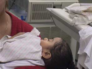
Infant put to sleep by mother. |
|
 |
|
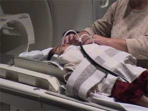
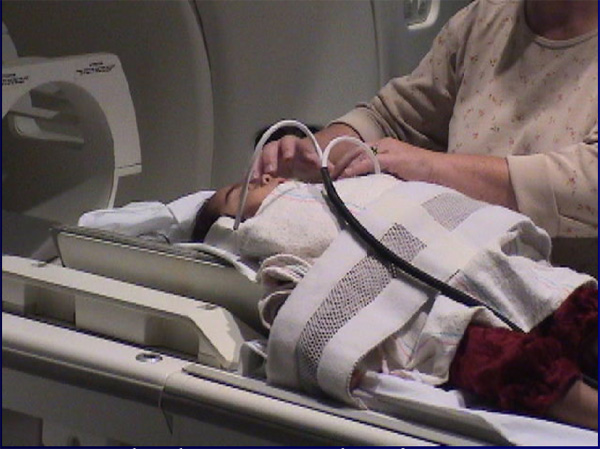
Headphones being attached. |
|
 |
|
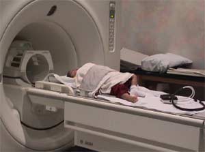
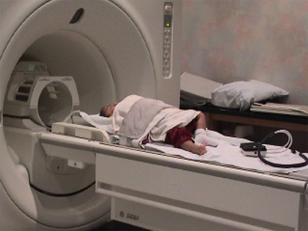
Infant nicely tucked, at foot of magnet. |
|
 |
|
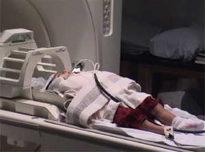
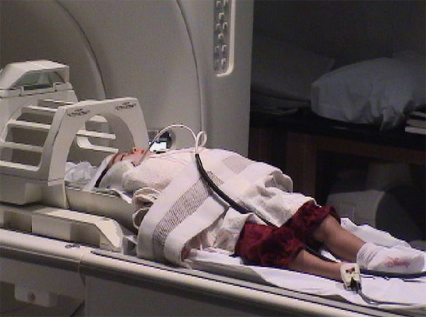
Infant inserted into head coil and magnet. |
Play audio/video clip:
When
presented with an auditory stimulus the auditory cortex "lights up" in an fMRI
study.(.avi file, 8 seconds)
Example of "motherese" speech (.wav
file, 9 seconds)
View related abstract:
fMRI of Auditory Activation from Language Stimulation in Neonates
# # #
 PDF
PDF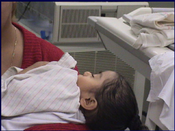