RSNA Press Release
- Hip pain in golfers may mean injury to the cartilage within the hip joint (labrum) — not arthritis, as is often diagnosed.
- Tears and detachments of the labrum can be corrected with arthroscopic surgery, a less invasive alternative to hip replacement.
- Professional golfers average three injuries from the sport and amateurs average two, with a growing awareness on hip injuries.
Hip Cartilage is Newest Achilles Heel for Golfers
Released: December 1, 2003
|
Media Contacts: |
Heather Babiar or Maureen Morley | (630) 590-7762 |
|
Heather Babiar |
Maureen Morley (630) 590-7754 mmorley@rsna.org |
CHICAGO — Shoulders, elbows and wrists aren't the only vulnerable joints for golfers. Add hips to the list, specifically injury to the labrum cartilage, a condition often diagnosed as arthritis or muscle strain.
"Ten years ago, labral tears were infrequently diagnosed," said Derek R. Armfield, M.D., assistant professor in the division of musculoskeletal radiology at the University of Pittsburgh Medical Center (UPMC). "The ability to detect labral injuries using magnetic resonance imaging has increased significantly in the last 10 years. At the same time, arthroscopic interventions have been developed to treat these injuries without subjecting the patient to major surgery."
Dr. Armfield and colleagues studied eight professional golfers suffering from hip pain who underwent a physical examination, magnetic resonance (MR) imaging and subsequent hip arthroscopy. He presented his findings today at the 89th Scientific Assembly and Annual Meeting of the Radiological Society of North America (RSNA).
The MR images demonstrated injuries to each patient's labrum, the ligament-like cartilage that contains nerves and lines the socket of the hip. It is believed that these tears can damage the adjacent cartilage and may lead to arthritis. Labral abnormalities included tears and detachments, and were highly correlated with abnormal results on arthroscopy exams, making MR imaging a good first step for diagnosing hip pain, Dr. Armfield said. All eight professional golfers were successfully treated with arthroscopic surgery.
"In the past, patients with hip pain were often diagnosed with arthritis, and the only treatment option offered was a total hip replacement somewhere down the road," said one of the study's co-authors, Douglas D. Robertson, M.D., Ph.D. "Today, patients living with hip pain can not only be diagnosed and treated, they also can return to their previous level of activity."
Chronic pain is common in golfers at all playing levels, Dr. Armfield said. According to a study of German golfers published in the June 2003 issue of the American Journal of Sports Medicine, professional golfers average three injuries from the sport and amateurs average two.
"This is a wake-up call that hip injuries are not uncommon for golfers
and hip pain should not be ignored," Dr. Robertson said.
Additional authors of the study being presented are Jeffrey D. Towers, M.D.,
Hal D. Martin, M.D., Scott M. Lephart, Ph.D., and Marc J. Philippon, M.D.
RSNA is an association of more than 35,000 radiologists, radiation oncologists
and related scientists committed to promoting excellence in radiology through
education and by fostering research, with the ultimate goal of improving patient
care. The Society is based in Oak Brook, Ill.
 |
|
|
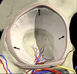
Side view of the horseshoe shaped hip socket with surrounding cartilage lining called the labrum (grey rim marked by arrows) which can tear and cause pain (reprinted with permission, Primal Pictures, Ltd). |
|
 |
|
|
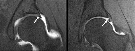
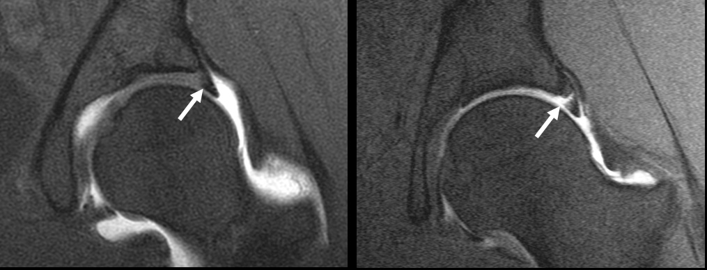
Frontal view of hip MRI showing normal appearance of labrum on the left (well defined dark triangle attached to the rim of the hip socket) as compared to abnormal torn labrum detached from the hip socket (right). |
|
 |
|
|
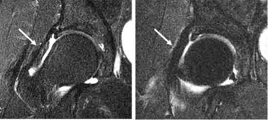
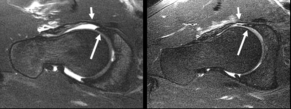
Cross sectional view of the normal triagular shaped labrum (long arrow) in relation to the hip along with normal appearance of frontal (anterior) capsule which helps keep the round femoral head in position. Abnormal labrum on right is thin and irregular (long arrow), the capsule is smaller as well (short arrow). |
|
 |
|
|
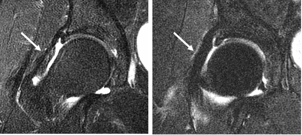
Frontal view of the hip shows thin weak capsule on the left and surgically reinforced capsule on the right which helps to keep the hip in place. |
|
 |
|
|
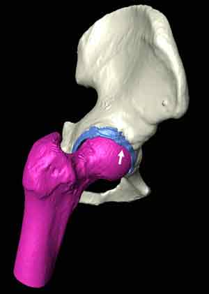
3-D model of the hip (made from CT scan) simulates position of the hip joint before golf ball impact. The arrow shows the femoral head (purple ball) colliding with the labrum (lining of the hip socket in blue) which may cause painful tears of the labrum. |
View related abstract:
# # #
 PDF
PDF