Black Patients Face Delays in Alzheimer’s Diagnosis
Released: November 27, 2023
At A Glance
- Researchers found that Black patients were imaged for cognitive impairment at an older age than white patients and were less likely to undergo MRI, the optimal exam for cognitive impairment and Alzheimer’s disease diagnosis.
- The average age at imaging for Black patients was 72.5 years, compared with 67.8 years for white patients and 66.5 years for Hispanics.
- Only 50.9% of Black patients underwent MRI testing, compared to 60% of whites and 67% of Hispanics.
- RSNA Media Relations
1-630-590-7762
media@rsna.org - Linda Brooks
1-630-590-7738
lbrooks@rsna.org - Imani Harris
1-630-481-1009
iharris@rsna.org
CHICAGO — Black patients underwent medical imaging for cognitive impairment years later than white and Hispanic patients and were less frequently tested with MRI, according to research being presented this week at the annual meeting of the Radiological Society of North America (RSNA).

Previous studies have shown that Black patients are at increased risk of Alzheimer’s disease and other types of dementia. They are less likely to have a diagnosis and are diagnosed at a more advanced stage of disease compared to white patients.
Medical imaging—ideally with MRI—plays an increasingly important role in the diagnostic work-up of cognitive impairment. However, it is unknown how disparities in imaging access may lead to these delays in cognitive impairment diagnoses.
“If disparity in obtaining access to neuroimaging is one possible barrier that delays diagnosis, it is important to identify this and figure out possible solutions to benefit these patients and prevent a delayed diagnosis,” said study lead author Joshua Wibecan, M.D., radiology resident at Boston Medical Center in Boston, Massachusetts.
Drawing from four years of data, Dr. Wibecan and colleagues studied imaging disparities at their safety net academic medical center. A safety net medical center provides health care for people regardless of their insurance status or ability to pay.
The researchers identified all outpatient CTs of the head, CT angiographies of the head and MRI brain examinations performed for cognitive impairment. They obtained patient self-identified race from the Boston Medical Center Clinical Data Warehouse for Research.
Self-identified Black/African American patients were imaged for cognitive impairment at an older age and were less frequently imaged for cognitive impairment with MRI. While CT and MRI can both be useful in detection of cognitive impairment and dementia diagnoses, MRI provides much more detail about brain abnormalities.
The average age at imaging for cognitive impairment among the groups with Black patients was 72.5 years, compared with 67.8 years for white patients, 66.5 years for Hispanics and 66.7 years for the Other group. Only 50.9% of Black patients underwent MRI testing for cognitive impairment, compared to 60% of white patients, 67% of Hispanics and 68.2% in the Other group.
“Our study demonstrates two main findings,” Dr. Wibecan said. “First, Black patients who received MRI or CT for cognitive impairment were significantly older than patients from other races. Second, Black patients were significantly less likely to be imaged with MRI, the optimal type of imaging for cognitive impairment, as opposed to CT.”
Early imaging evaluation is important to identify treatable causes of cognitive impairment, such as tumors, bleeding or swelling within the brain. Additionally, new treatments have recently become available for Alzheimer’s disease that can potentially slow the rate of decline. Earlier diagnosis may, therefore, lead to early treatment and a longer period of better cognitive function.
“As treatment for Alzheimer’s Disease improves, it will be even more important to identify patients at early stages of disease for optimal treatment,” Dr. Wibecan said.
Further research is needed, Dr. Wibecan said, to understand why there was a significant difference in the types of imaging exams ordered for the workup of cognitive impairment across racial groups.
Chad W. Farris, M.D., Ph.D., neuroradiologist from Boston Medical Center and assistant professor of radiology at Boston University Chobanian & Avedisian School of Medicine, co-authored the study.
Note: Copies of RSNA 2023 news releases and electronic images will be available online at RSNA.org/press23.
RSNA is an association of radiologists, radiation oncologists, medical physicists and related scientists promoting excellence in patient care and health care delivery through education, research and technologic innovation. The Society is based in Oak Brook, Illinois. (RSNA.org)
Editor’s note: The data in these releases may differ from those in the published abstract and those actually presented at the meeting, as researchers continue to update their data right up until the meeting. To ensure you are using the most up-to-date information, please call the RSNA Newsroom at 1-312-791-6610.
For patient-friendly information on brain MRI and CT, visit RadiologyInfo.org.
Video (MP4):

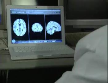
Video 1. Joshua Wibecan, M.D., discusses his research on how Black patients face delays in Alzheimer’s diagnosis.
Download MP4
(Right-click and Save As)
Images (JPG, TIF):
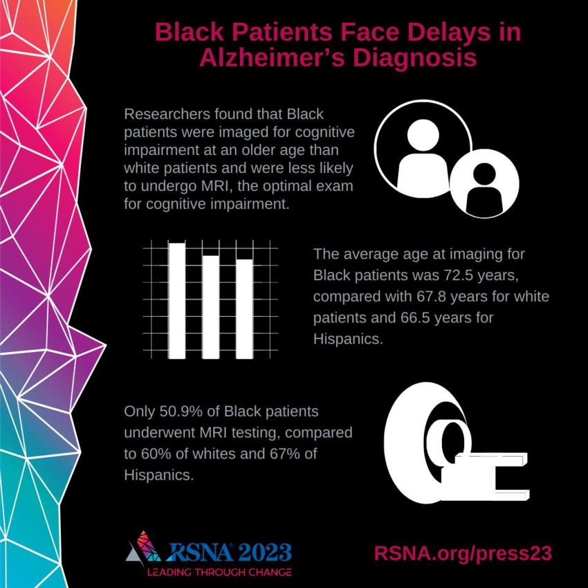
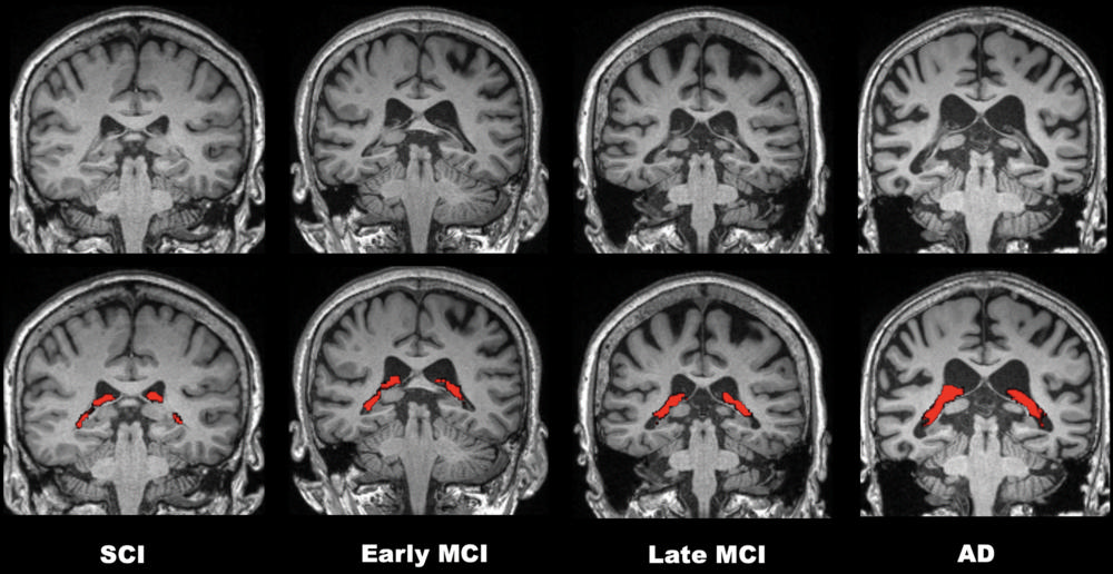
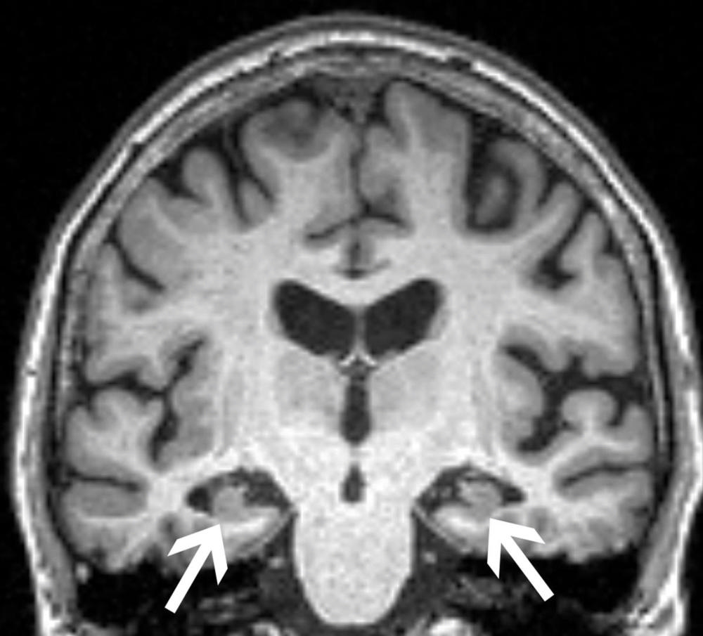
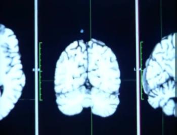
Figure 2. Brain MRI in patient with mild cognitive impairment.
High-res (TIF) version
(Right-click and Save As)
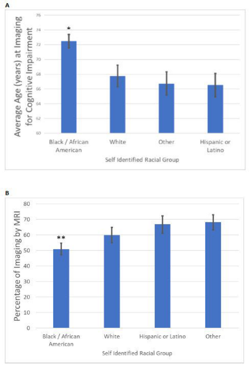
Figure 3. (A) Average age in years at imaging for cognitive impairment by self-identified racial group. (B) Percentage of imaging for cognitive impairment performed by MRI by self-identified racial group.
High-res (TIF) version
(Right-click and Save As)
Additional Resources