Soccer Heading Linked to Measurable Decline in Brain Function
Released: November 28, 2023
At A Glance
- Soccer heading by young adult amateur players over a two-year period is linked to measurable decline in the microstructure and function of the brain.
- Researchers found that high levels of heading over the two-year period were associated with changes in brain microstructure similar to findings seen in mild traumatic brain injury.
- A brain region called the gray and white matter interface was blunted in proportion to high repetitive head impact exposure.
- RSNA Media Relations
1-630-590-7762
media@rsna.org - Linda Brooks
1-630-590-7738
lbrooks@rsna.org - Imani Harris
1-630-481-1009
iharris@rsna.org
CHICAGO — New research being presented this week at the annual meeting of the Radiological Society of North America (RSNA) links soccer heading – where players hit the ball with their head – to a measurable decline in the microstructure and function of the brain over a two-year period.

“There is enormous worldwide concern for brain injury in general and in the potential for soccer heading to cause long-term adverse brain effects in particular,” said senior author Michael L. Lipton, M.D., Ph.D., professor of radiology at Columbia University’s Vagelos College of Physicians and Surgeons and affiliate professor of biomedical engineering at Columbia University. “A large part of this concern relates to the potential for changes in young adulthood to confer risk for neurodegeneration and dementia later in life.”
While previous research has examined adverse effects on the brain related to soccer heading at a single point in time, this new study looked at brain changes over two years.
The study included 148 young adult amateur soccer players (mean age 27, 26% women). The research team developed a specialized questionnaire for players to determine how often they hit the soccer ball with their head.

“When we first started, there was no method for assessing the number of head impacts a player experienced,” Dr. Lipton said. “So, we developed a structured, epidemiological questionnaire that has been validated in multiple studies.”
The questionnaire consists of a series of questions about how often an individual plays, practices and heads the ball, and in what type of situations. Two-year heading exposure was categorized as low, moderate or high.
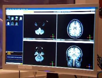
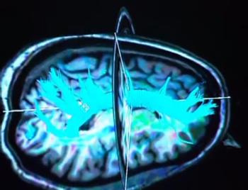
The players were assessed for verbal learning and memory and underwent diffusion tensor imaging (DTI), an MRI technique, at the time of enrollment and two years later. DTI characterizes the microstructure of the brain by tracking the microscopic movement of water molecules through the tissue.
Compared to the baseline test results, the high-heading group (over 1,500 headers in two years) demonstrated an increase of diffusivity in frontal white matter regions, and a decrease of orientation dispersion index (a measure of brain organization) in certain brain regions after two years of heading exposure. The analysis adjusted for variables including age, sex, education and concussion history.
“Our analysis found that high levels of heading over the two-year period were associated with changes in brain microstructure similar to findings seen in mild traumatic brain injuries,” Dr. Lipton said. “High levels of heading were also associated with a decline in verbal learning performance. This is the first study to show a change of brain structure over the long term related to sub-concussive head impacts in soccer.”
Dr. Lipton and colleagues also presented another study today in which they used DTI to investigate the association between repetitive head impacts from soccer heading and verbal learning performance.

For the second study, researchers analyzed heading over 12 months prior to DTI and verbal learning performance testing in 353 amateur soccer players (age 18-53, 27% female). Unlike previous research that has focused on deep white matter regions, this study employed a new technique, using DTI parameters to evaluate the integrity of the interface between the brain’s gray and white matter closer to the skull.
“Importantly, our new approach addresses a brain region that is susceptible to injury but has been neglected due to limitations of existing methods,” Dr. Lipton said. “Application of this technique has potential to disclose the extent of injury from repetitive heading, but also from concussion and traumatic brain injury to an extent not previously possible.”
The researchers found that the normally sharp gray matter-white matter interface was blunted in proportion to high repetitive head impact exposure.
“We used DTI to assess the sharpness of the transition from gray matter to white matter,” Dr. Lipton said. “In various brain disorders, what is typically a sharp distinction between these two brain tissues becomes a more gradual, or fuzzier transition.”
He added that gray matter-white matter interface integrity may play a causal role in the adverse association between repetitive head impacts and cognitive performance.
“These findings add to the ongoing conversation and contentious debate as to whether soccer heading is benign or confers significant risk,” he said.
Co-authors on the first study are Molly F. Charney, M.D., Kenny Ye, Ph.D., Roman Fleysher, Ph.D., Liane E. Hunter, M.D., Ph.D., Shimon Garrel, B.S., Bluyé Demessie, A.B., M.S., Joan Y. Song, B.S.E., M.S., Molly E. Zimmerman, Ph.D., Walter F. Stewart, Ph.D., Mimi Kim, Sc.D., and Richard B. Lipton, M.D.
Co-authors on the second study are Joan Y. Song, B.S.E., M.S., and Roman Fleysher, Ph.D.
Note: Copies of RSNA 2023 news releases and electronic images will be available online at RSNA.org/press23.
RSNA is an association of radiologists, radiation oncologists, medical physicists and related scientists promoting excellence in patient care and health care delivery through education, research and technologic innovation. The Society is based in Oak Brook, Illinois. (RSNA.org)
Editor’s note: The data in these releases may differ from those in the published abstract and those actually presented at the meeting, as researchers continue to update their data right up until the meeting. To ensure you are using the most up-to-date information, please call the RSNA Newsroom at 1-312-791-6610.
For patient-friendly information on brain imaging visit RadiologyInfo.org.
Video (MP4):
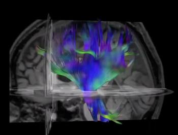
Video 1. Diffusion tensor imaging, an MRI technique, of the brain.
Download MP4
(Right-click and Save As)
Video 6. Diffusion tensor imaging, an MRI technique, of the brain.
Download MP4
(Right-click and Save As)
Images (JPG, TIF):
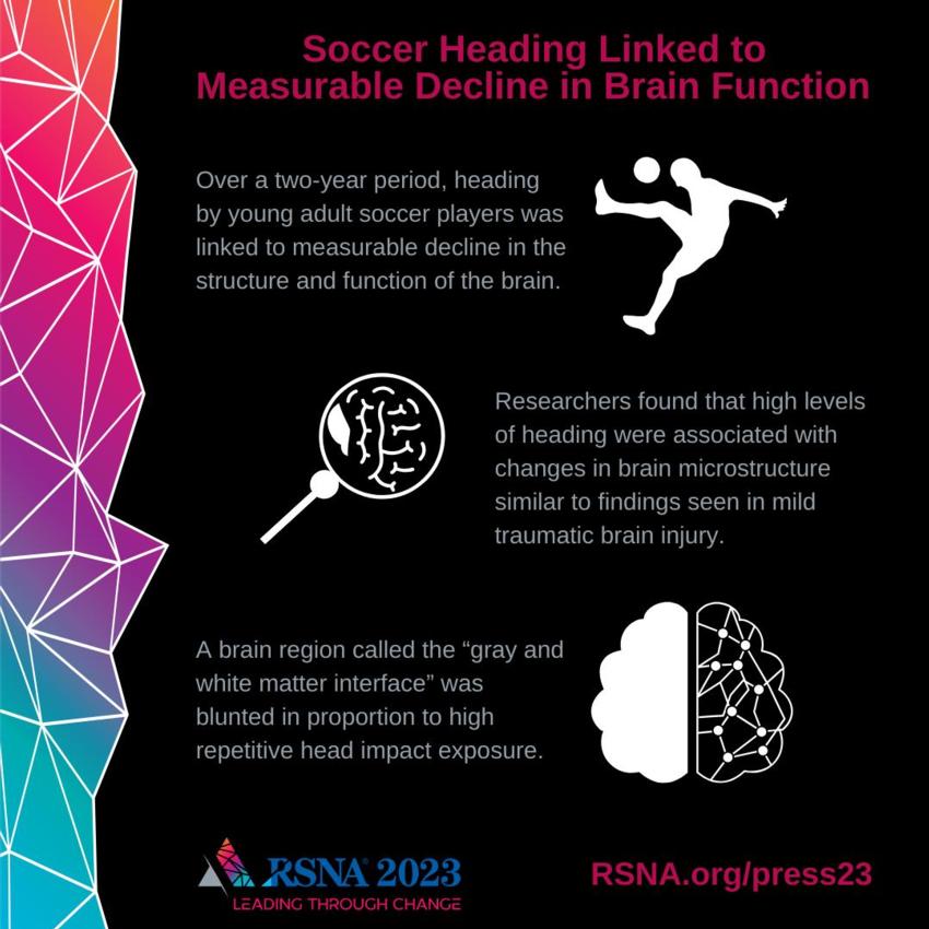
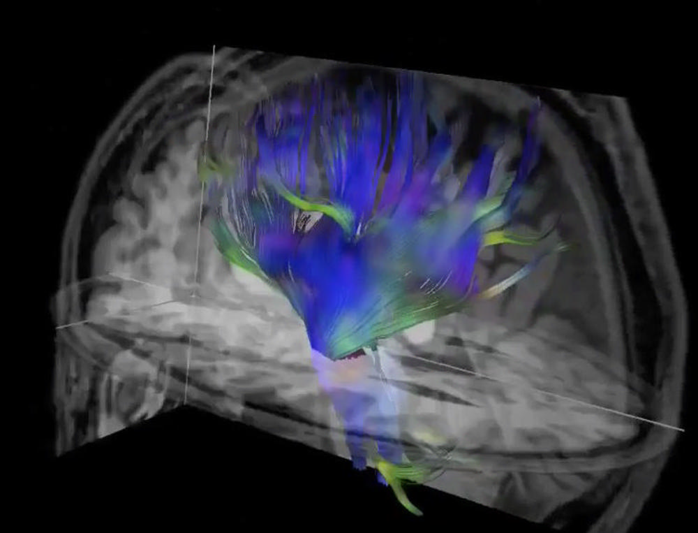
Figure 1. Diffusion tensor imaging, an MRI technique, of the brain.
High-res (TIF) version
(Right-click and Save As)
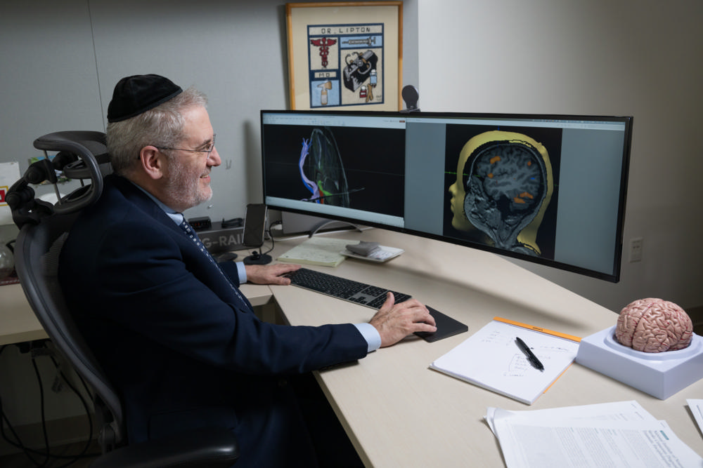
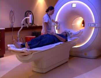
Figure 2. Dr. Michael Lipton reviewing brain images.
High-res (TIF) version
(Right-click and Save As)
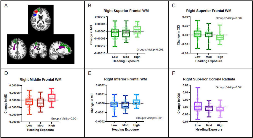
Figure 3. (A) Regions of interest are highlighted on an example T1 brain (green = right superior frontal WM, red = right middle frontal WM, blue = right inferior frontal WM. Purple = right superior corona radiata). (B-F) Change in DTI and NODDI measures (MD and ODI) by heading group in four regions of interest.
High-res (TIF) version
(Right-click and Save As)
Additional Resources