3-D Mammography Improves Cancer Detection in Dense Breasts
Released: December 02, 2014
At A Glance
- A study of more than 25,000 women has found that digital tomosynthesis significantly increases the cancer detection rate in dense breast tissue.
- Using digital mammography plus tomosynthesis, researchers detected 80 percent of 132 cancers in women with dense breasts, compared to only 59 percent for mammography alone.
- Tomosynthesis provides 3-D views of the breast.
- RSNA Media Relations
1-630-590-7762
media@rsna.org - Maureen Morley
1-630-590-7754
mmorley@rsna.org - Linda Brooks
1-630-590-7738
lbrooks@rsna.org
CHICAGO – A major new study being presented at the annual meeting of the Radiological Society of North America (RSNA) has found that digital breast tomosynthesis, also known as 3-D mammography, has the potential to significantly increase the cancer detection rate in mammography screening of women with dense breasts.

Breasts are considered dense if they have a lot of fibrous or glandular tissue but not much fatty tissue. Research has shown that dense breasts are more likely to develop cancer, a problem compounded by the fact that cancer in dense breasts can be difficult to detect on mammograms.
Other imaging modalities like ultrasound and MRI are often used to help find cancers that can't be seen on mammograms, but both modalities have higher rates of false-positive findings, which are suspicious findings that turn out not to be cancer. This higher false-positive rate often results in more tests and unnecessary biopsies, making MRI and ultrasound expensive to implement in high-volume screening programs, according to study lead author Per Skaane, M.D., Ph.D., from the Department of Radiology at Oslo University Hospital in Oslo, Norway.
Dr. Skaane and colleagues have been studying tomosynthesis as a promising breast cancer screening option that addresses some of the limitations of mammography by providing 3-D views of the breast.
"Tomosynthesis could be regarded as an improvement of mammography and would be much easier than MRI or ultrasound to implement in organized screening programs," Dr. Skaane said. "So the intention of our study was to see if tomosynthesis really would significantly increase the cancer detection rate in a population-based mammography screening program."
The researchers compared cancer detection using full-field digital mammography (FFDM) versus FFDM plus digital breast tomosynthesis in 25,547 women between the ages of 50 and 69. Breast density was classified based on the American College of Radiology's Breast Imaging-Reporting and Data System (BI-RADS). The BI-RADS breast density scale runs from 1 to 4, with 1 being the least dense and 4 being the most dense.
There were 257 malignancies detected on FFDM and a combination of FFDM and tomosynthesis in the study group, including 105 in the density 2 group and 110 in density 3. Of the 257 cancers, 211, or 82 percent were detected with FFDM plus tomosynthesis, a significant improvement over the 163, or 63 percent, detected with FFDM alone.
FFDM plus tomosynthesis pinpointed 80 percent of the 132 cancer cases in women with dense breasts, compared to only 59 percent for FFDM alone.
"Our findings are extremely promising, showing an overall relative increase in the cancer detection rate of about 30 percent," Dr. Skaane said. "Stratifying the results on invasive cancers only, the relative increase in cancer detection was about 40 percent."
Tomosynthesis not only improved the cancer detection rate in women with dense breasts, it also helped increase detection for women in the "fatty breast" BI-RADS categories. The addition of tomosynthesis to FFDM improved the cancer detection rate from 68 percent to 84 percent in women with BI-RADS density 1 or 2 breasts.
"Our results show that implementation of tomosynthesis might indicate a new era in breast cancer screening," Dr. Skaane said.
Co-authors on the study are Bjorn Helge Osteras, M.Sc., Ellen B. Eben, M.D., and Randi Gullien, R.T.
Note: Copies of RSNA 2014 news releases and electronic images will be available online at RSNA.org/press14 beginning Monday, Dec. 1.
RSNA is an association of more than 54,000 radiologists, radiation oncologists, medical physicists and related scientists, promoting excellence in patient care and health care delivery through education, research and technologic innovation. The Society is based in Oak Brook, Ill. (RSNA.org)
Editor's note: The data in these releases may differ from those in the published abstract and those actually presented at the meeting, as researchers continue to update their data right up until the meeting. To ensure you are using the most up-to-date information, please call the RSNA Newsroom at 1-312-791-6610.
For patient-friendly information on breast imaging, visit RadiologyInfo.org.
Images (.JPG and .TIF format)
and Video clips (.mp4 format)
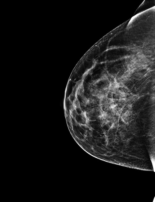
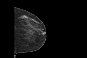
Figure 1. 46-yr-old woman 2-D full-field digital mammography view of breast.
High-res (TIF) version
(Right-click and Save As)
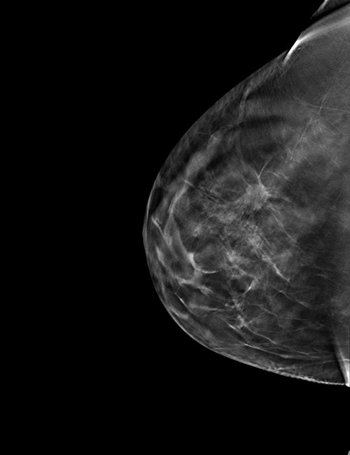
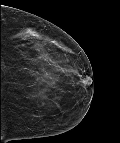
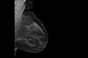
Figure 2. 46-yr-old woman 3-D digital tomosynthesis view of breast.
High-res (TIF) version
(Right-click and Save As)
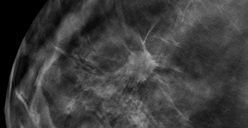
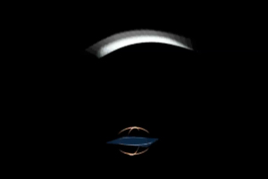
Figure 3. Spiculated mass on 3-D tomosynthesis image.
High-res (TIF) version
(Right-click and Save As)

Figure 4. A 46-year-old woman’s routine screening mammogram: 2-D mammogram is essentially negative. 3-D Tomosynthesis images reveal a 15 mm spiculated mass between slices 38-48, diagnosed as invasive ductal carcinoma, grade 2.
High-res (TIF) version
(Right-click and Save As)
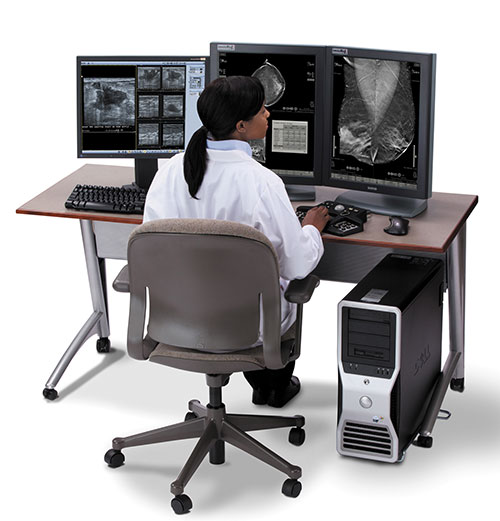
Figure 5. A radiologist reading a 3-D mammogram on a diagnostic workstation.
High-res (TIF) version
(Right-click and Save As)
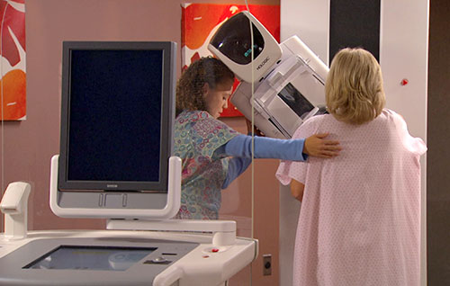
Figure 6. A patient being imaged with the 3-D mammography system.
High-res (TIF) version
(Right-click and Save As)

Video 3. Dr. Per Skaane discusses digital tomosynthesis-Part 1
Download.mp4
(Right-click and Save As)

Video 4. Dr. Per Skaane discusses digital tomosynthesis-Part 2
Download.mp4
(Right-click and Save As)