Researchers Use AI to Track Cognitive Deviation in Aging Brains
Released: June 23, 2021
At A Glance
- Researchers built a model to track deviation from healthy brain aging.
- The brain age prediction model was trained based on the MR images of 974 healthy adults.
- The model has potential for early detection of cognitive impairment at an individual level.
- RSNA Media Relations
1-630-590-7762
media@rsna.org - Linda Brooks
1-630-590-7738
lbrooks@rsna.org - Katherine Anderson
1-630-491-1009
kanderson@rsna.org
OAK BROOK, Ill. — Researchers have developed an artificial intelligence (AI)-based brain age prediction model to quantify deviations from a healthy brain-aging trajectory in patients with mild cognitive impairment, according to a study published in Radiology: Artificial Intelligence. The model has the potential to aid in early detection of cognitive impairment at an individual level.
Amnestic mild cognitive impairment (aMCI) is a transition phase from normal aging to Alzheimer’s disease (AD). People with aMCI have memory deficits that are more serious than normal for their age and education, but not severe enough to affect daily function.
For the study, Ni Shu, Ph.D., from State Key Laboratory of Cognitive Neuroscience and Learning, Beijing Normal University, in Beijing, China, and colleagues used a machine learning approach to train a brain age prediction model based on the T1-weighted MR images of 974 healthy adults aged from 49.3 to 95.4 years. The trained model was applied to estimate the predicted age difference (predicted age vs. actual age) of aMCI patients in the Beijing Aging Brain Rejuvenation Initiative (616 healthy controls and 80 aMCI patients) and the Alzheimer’s Disease Neuroimaging Initiative (589 healthy controls and 144 aMCI patients) datasets.
The researchers also examined the associations between the predicted age difference and cognitive impairment, genetic risk factors, pathological biomarkers of AD, and clinical progression in aMCI patients.
The results showed that aMCI patients had brain-aging trajectories distinct from the typical normal aging trajectory, and the proposed brain age prediction model could quantify individual deviations from the typical normal aging trajectory in these patients. The predicted age difference was significantly associated with individual cognitive impairment of aMCI patients in several domains, specifically including memory, attention and executive function.
“The predictive model we generated was highly accurate at estimating chronological age in healthy participants based on only the appearance of MRI scans,” the researchers wrote. “In contrast, for aMCI, the model estimated brain age to be greater than 2.7 years older on average than the patient’s chronological age.”
The model further showed that progressive aMCI patients exhibit more deviations from typical normal aging than stable aMCI patients, and the use of the predicted age difference score along with other AD-specific biomarkers could better predict the progression of aMCI. Apolipoprotein E (APOE) ε4 carriers showed larger predicted age differences than non-carriers, and amyloid-positive patients showed larger predicted age differences than amyloid-negative patients.
Combining the predicted age difference with other biomarkers of AD showed the best performance in differentiating progressive aMCI from stable aMCI.
“This work indicates that predicted age difference has the potential to be a robust, reliable and computerized biomarker for early diagnosis of cognitive impairment and monitoring response to treatment,” the authors concluded.
“Accelerated Brain Aging in Amnestic Mild Cognitive Impairment: Relationships with Individual Cognitive Decline, Risk Factors for Alzheimer Disease and Clinical Progression.” Weijie Huang, Xin Li, He Li, Wenxiao Wang, Kewei Chen, Kai Xu, Junying Zhang, Yaojing Chen, Dongfeng Wei, Ni Shu, Ph.D., and Zhanjun Zhang.
Radiology: Artificial Intelligence is edited by Charles E. Kahn Jr., M.D., M.S., Perelman School of Medicine at the University of Pennsylvania, and owned and published by the Radiological Society of North America, Inc. (https://pubs.rsna.org/journal/ai)
RSNA is an association of radiologists, radiation oncologists, medical physicists and related scientists promoting excellence in patient care and health care delivery through education, research and technologic innovation. The Society is based in Oak Brook, Illinois. (RSNA.org)
For information on brain MRI and Alzheimer’s Disease, visit RadiologyInfo.org.
Press Resources:
Images (JPG, TIF):

Figure 1. Flowchart showing the framework of the brain age prediction model. A, The imaging data were split into training and test datasets. The training dataset consisted of structural magnetic resonance imaging data from 974 healthy individuals, whereas the test dataset included data from 2 groups, 231 healthy controls and 224 aMCI subjects. B, A Conventional Statistical Parametric Mapping structural preprocessing pipeline was used to generate GMV maps in the MNI space. C, The intensity values from the GMV maps were extracted and concatenated to create a feature matrix that was then cleaned and normalized. D, The best elastic net model was obtained by performing supervised learning on the training dataset. To optimize the hyperparameters, a grid search was performed. E, The test dataset was input into the trained model. An age was predicted for every participant included in the test dataset. The PAD scores were calculated by subtracting the participant’s chronological age from his or her predicted age. aMCI = amnestic mild cognitive impairment, GMV = gray matter volume, MNI = Montreal Neurologic Institute, Dartel = Diffeomorphic Anatomic Registrations Through Exponentiated Lie Algebra, PAD = predicted age difference.
High-res (TIF) version
(Right-click and Save As)
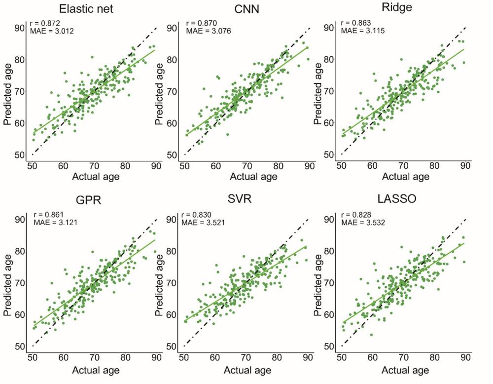
Figure 2. Performance of different machine learning approaches in predicting the chronological age of healthy controls in the test dataset. Scatterplot of the actual age and brain age predicted by different machine learning methods. CNN = Convolutional neural network, GPR = Gaussian process regression, SVR = support vector regression, LASSO = least absolute shrinkage and selection operator.
High-res (TIF) version
(Right-click and Save As)
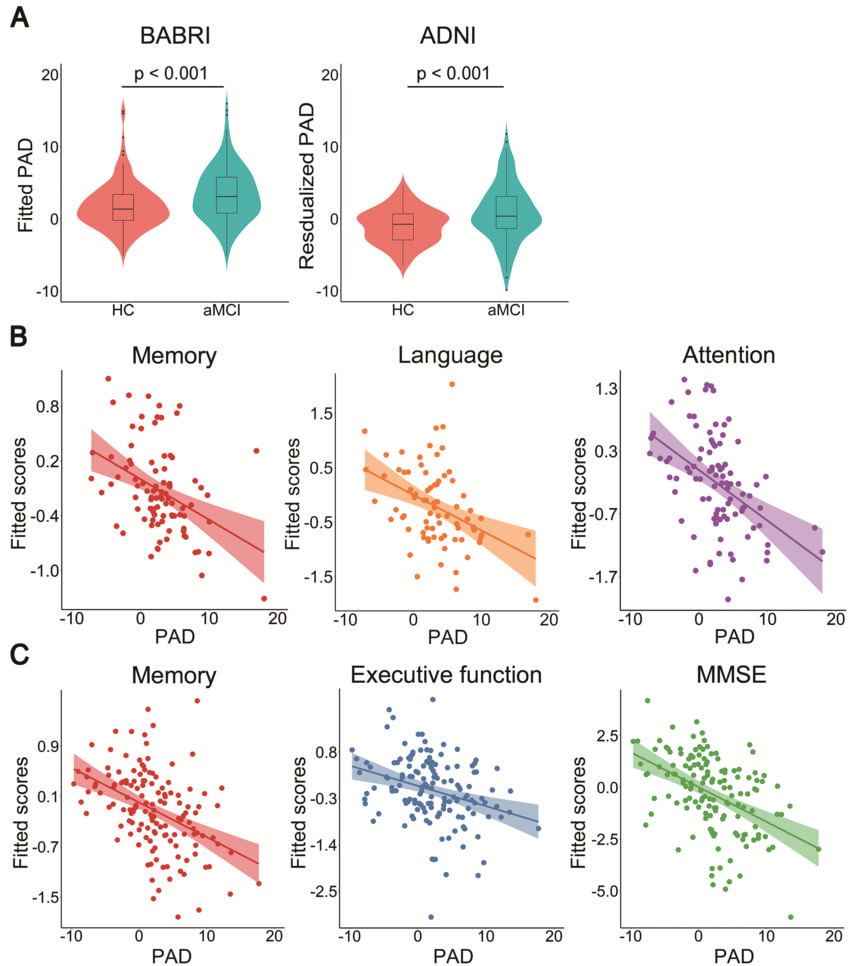
Figure 3. Relationship between the PAD and cognitive impairment in aMCI patients. A, The group differences in PAD scores between the HCs and aMCI patients included in the BABRI (T = 4.17) and ADNI (W = 5298) datasets. B, There was a significant correlation between the PAD and memory, language and attention scores in the aMCI patients included in the BABRI dataset. C, There was a significant correlation between the PAD and memory, executive function and MMSE scores in the aMCI patients included in the ADNI dataset. BABRI = Beijing Ageing Brain Rejuvenation Initiative, ADNI = Alzheimer’s Disease Neuroimaging Initiative, aMCI = amnestic mild cognitive impairment, PAD = predicted age difference, MMSE = Mini-Mental Status Examination.
High-res (TIF) version
(Right-click and Save As)
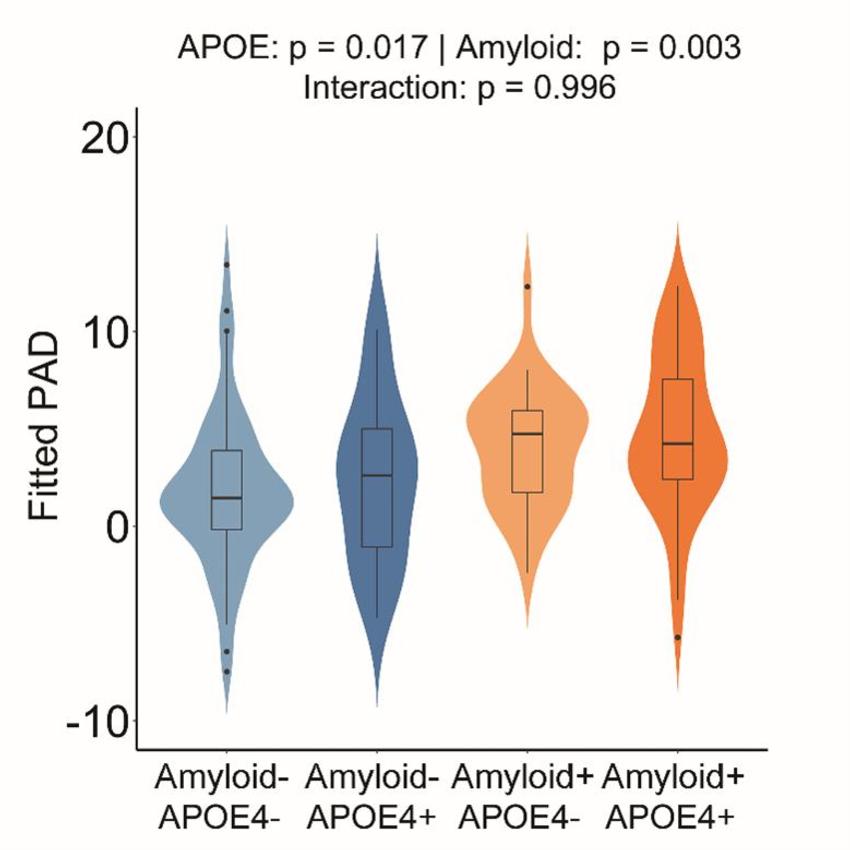
Figure 4. Effects of different AD risk factors on the PAD in aMCI patients. The main effects of APOE and amyloid status on the PAD in aMCI patients were statistically significant, while the amyloid APOE status interaction effect on the PAD was not significant. There were 54 APOE ε4 noncarriers with amyloid-negative status, 28 APOE ε4 noncarriers with amyloid-positive status, 17 APOE ε4 carriers with amyloid-negative status and 45 APOE ε4 carriers with amyloid-positive status. APOE = apolipoprotein E, APOE4- = APOE ε4 noncarriers, APOE4+ = APOE ε4 carriers, Amyloid- = amyloid-negative patients, Amyloid+ = amyloid positive patients.
High-res (TIF) version
(Right-click and Save As)
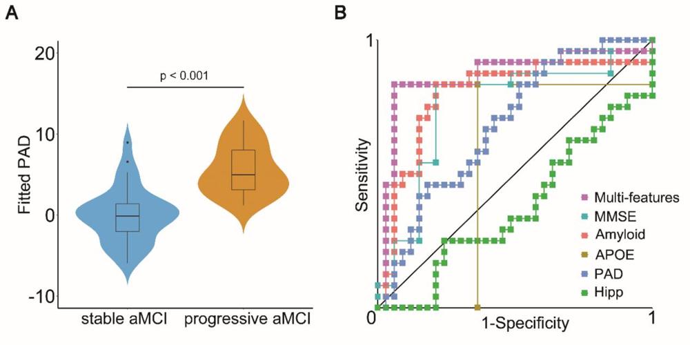
Figure 5. Clinical progression prediction with the PAD in aMCI patients. A, The progressive aMCI group showed a larger PAD than the stable aMCI group. B, The PAD at baseline combined with the MMSE score, amyloid SUVr value and APOE allele status outperformed any single feature in discriminating progressive aMCI from stable aMCI. PAD = predicted age difference, MMSE = Mini-Mental Status Examination, Hipp = hippocampal volume, APOE = apolipoprotein E, Multifeatures = PAD, MMSE, amyloid and APOE allele status.
High-res (TIF) version
(Right-click and Save As)