Presence of Blood Clot Associated with Rapid Aortic Aneurysm Growth
Released: January 28, 2020
At A Glance
- Rapid, potentially dangerous growth in abdominal aortic aneurysms is associated with the presence of blood clots.
- The researchers used CT or MRI to assess 225 men with abdominal aortic aneurysm, and followed aneurysm growth for an average of 3 years.
- The findings could help identify which patients need more aggressive treatment and more frequent follow-up imaging.
- RSNA Media Relations
1-630-590-7762
media@rsna.org - Linda Brooks
1-630-590-7738
lbrooks@rsna.org - Dionna Arnold
1-630-590-7791
darnold@rsna.org
OAK BROOK, Ill. — The presence of a blood clot on the wall of the aorta in people with abdominal aortic aneurysms is associated with more rapid, potentially dangerous growth in the aneurysm, according to a major study published in the journal Radiology. Researchers said the findings could help identify which patients need more aggressive treatment and more frequent follow-up imaging after their initial diagnosis.

The aorta is the major artery that carries oxygenated blood from the heart. Abdominal aneurysms occur when a bulge forms in the portion of the artery that runs through the abdomen. About 200,000 people in the U.S. are diagnosed with the condition every year. Over time, the wall can weaken and rupture. Ruptured abdominal aortic aneurysm is the 10th leading cause of death for men over age 55.
Decisions to surgically repair the aneurysm are based on its diameter. Patients with aneurysms larger than 5.5 centimeters (cm) are normally referred for repair, while those smaller than 5.5 cm are most commonly monitored with imaging at regular intervals. Ultrasound and cross-sectional imaging with CT or MRI are commonly used.
However, this diameter-based management strategy has limitations, as a considerable number of small aneurysms rupture, according to the study's first author, Chengcheng Zhu, Ph.D., assistant researcher from the Department of Radiology and Biomedical Imaging at the University of California in San Francisco.
Dr. Zhu and colleagues focused their study on the intraluminal thrombus, a blood clot on the wall of the aorta at the location of the aneurysm. Intraluminal thrombi are present in the majority of aneurysms close to the repair threshold of 5.5 cm, and in a considerable number of smaller aneurysms. Despite their prevalence, the influence of these clots on abdominal aortic aneurysm growth and rupture risk is still not fully understood.
"Intraluminal thrombus could be a new marker for aneurysm growth," Dr. Zhu said.
The researchers used high resolution cross-sectional imaging with CT or MRI to assess 225 men with abdominal aortic aneurysm. Follow-up lasted, on average, more than 3 years.
Slightly more than half of patients had an intraluminal thrombus. The aneurysms of those with intraluminal thrombus were larger at baseline and grew by a rate of 2 millimeters (mm) per year, twice as fast as the 1 mm per year growth rate in people without intraluminal thrombus.
"An aneurysm with thrombus grows much faster than one without a thrombus," Dr. Zhu said. "Our study looked at a large number of patients with a relatively long follow-up to confirm that thrombus is a new risk factor that may be potentially reported by radiologists."
While the study did not examine the reasons why the presence of a thrombus had such a dramatic impact on aneurysms, Dr. Zhu noted that it likely has harmful biochemical effects on the vessel wall.
"When the vessel wall is covered with thrombus, the lack of oxygen weakens the wall of the vessel, making the aneurysm likely to grow faster and rupture," he said.
The findings suggest that imaging follow-up schedules may need to be adjusted for patients whose aneurysm has a thrombus. Current protocols for follow-up imaging are based on aneurysm size and do not account for the absence or presence of a thrombus.
"A patient at high risk may need closer monitoring," Dr. Zhu said. "If in patients with a thrombus the aneurysm grows twice as fast, then shortening the surveillance interval could be considered."
Dr. Zhu said more research is needed before thrombus detection is integrated into clinical protocols. For now, he said the findings provide another piece of information to use in determining the prognosis of patients with this potentially dangerous condition.
"Intraluminal Thrombus Predicts Rapid Growth of Abdominal Aortic Aneurysms." Collaborating with Dr. Zhu were Joseph R. Leach, M.D., Ph.D., Yuting Wang, M.D., Warren Gasper, M.D., David Saloner, Ph.D., and Michael D. Hope, M.D.
Radiology is edited by David A. Bluemke, M.D., Ph.D., University of Wisconsin School of Medicine and Public Health, Madison, Wis., and owned and published by the Radiological Society of North America, Inc. (http://radiology.rsna.org/)
RSNA is an association of radiologists, radiation oncologists, medical physicists and related scientists promoting excellence in patient care and health care delivery through education, research and technologic innovation. The Society is based in Oak Brook, Ill. (RSNA.org)
For patient-friendly information on abdominal aortic aneurysm and imaging, visit RadiologyInfo.org.
Images (JPG, TIF):
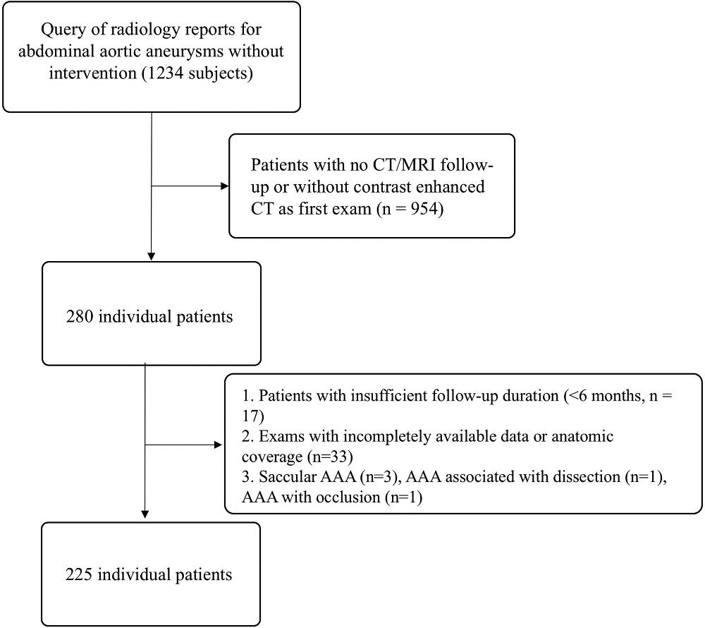
Figure 1. Patient selection flowchart. AAA = abdominal aortic aneurysm.
High-res (TIF) version
(Right-click and Save As)
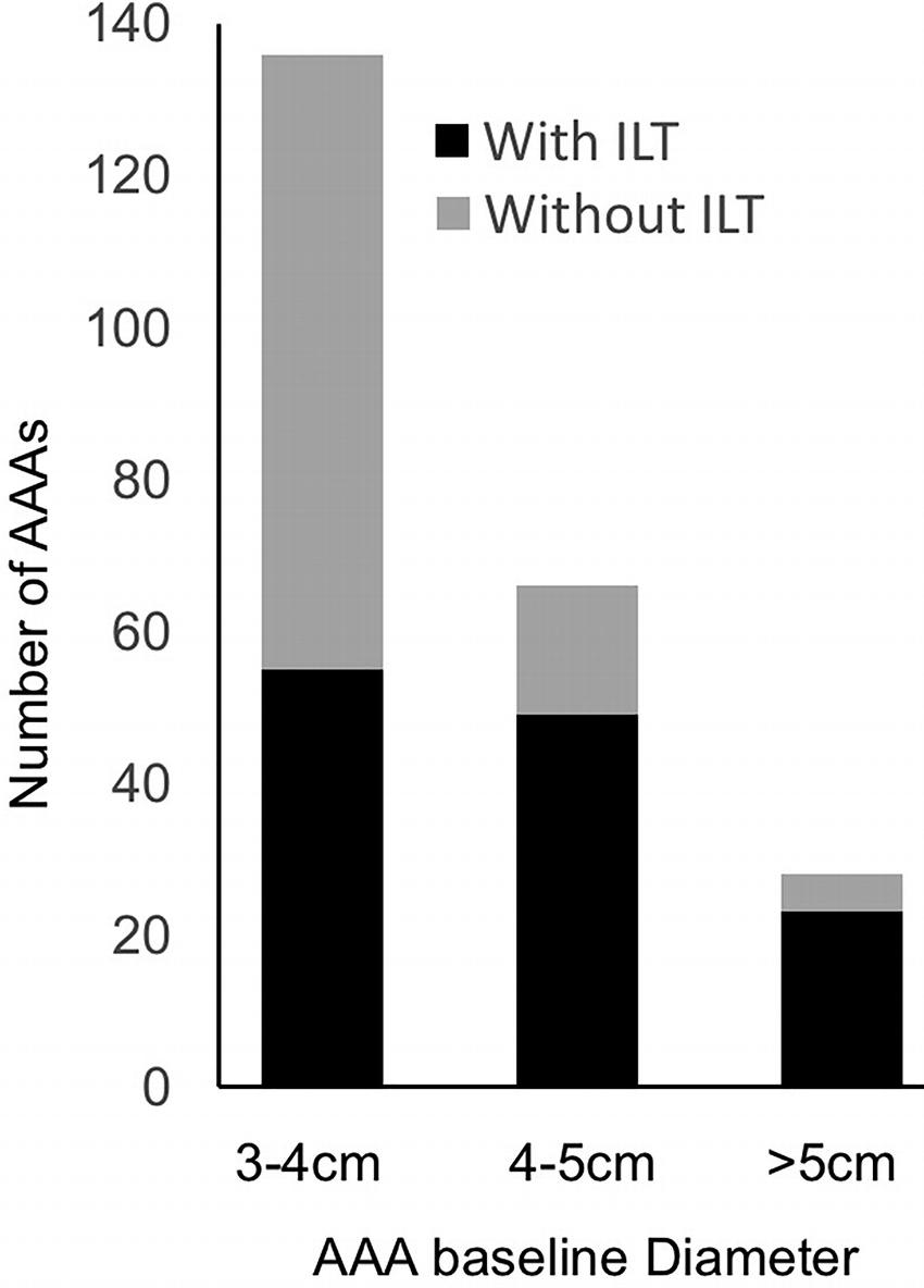
Figure 2. Distribution of intraluminal thrombus (ILT) across abdominal aortic aneurysm (AAA) diameter.
High-res (TIF) version
(Right-click and Save As)

Figure 3. Box-and-whisker plots showing the growth rate of 3–5-cm abdominal aortic aneurysm (AAA) with and without intraluminal thrombus (ILT). Left: The 3–4-cm group. Right: The 4–5-cm group. AAAs with ILT tend to grow 1.9 and 1.2 times faster than AAAs without ILT, even when their baseline diameters are comparable (median, 3.5 cm [interquartile range, 3.2–3.8 cm] vs 3.3 cm [interquartile range, 3.2 cm–3.6 cm]; P = .01; 4.4 cm [interquartile range, 4.2–4.7 cm] vs 4.2 cm [interquartile range, 4.2–4.3 cm]; P = .066). Actual points are shown for outliers.
High-res (TIF) version
(Right-click and Save As)
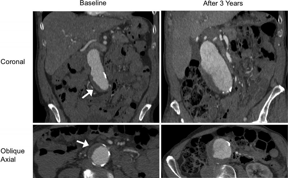
Figure 4. A patient (aged 85 years, male) with a fast-growing abdominal aortic aneurysm with intraluminal thrombus at baseline. Contrast-enhanced CT images (coronal and oblique axial planes) show that the aneurysm grew from 4.1 to 6.3 cm within 3 years at a growth rate of 7.3 mm/y. Arrow shows the intraluminal thrombus.
High-res (TIF) version
(Right-click and Save As)
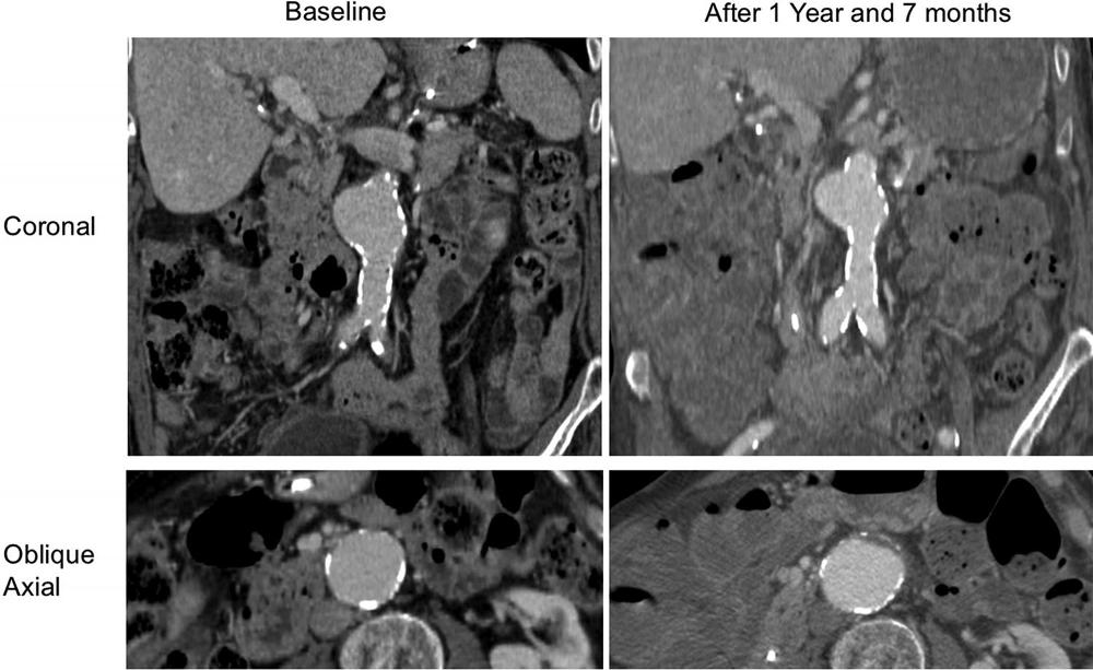
Figure 5. A patient (aged 88 years, male) with a slow-growing abdominal aortic aneurysm without intraluminal thrombus at baseline. Coronal and oblique axial contrast-enhanced CT images show that the aneurysm grew from 4.2 to 4.3 cm within 1 year and 7 months at a growth rate of 0.6 mm/y.
High-res (TIF) version
(Right-click and Save As)
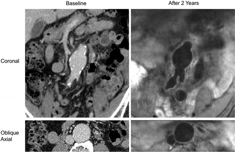
Figure 6. A patient (aged 83 years, male) who was followed up with black-blood MRI. The abdominal aortic aneurysm had no intraluminal thrombus at baseline or follow-up. Coronal and oblique axial contrast-enhanced CT images show that the aneurysm had a 4.0-cm diameter at baseline; 2 years later, black-blood MRI shows that the aneurysm grew to 4.2 cm at a growth rate of 1 mm/y.
High-res (TIF) version
(Right-click and Save As)