Novel Technique Accurately Assesses Cardiovascular Risks
Released: February 12, 2019
At A Glance
- A new technique for imaging carotid arteries may provide an earlier, more accurate assessment of cardiovascular disease risk.
- Volumetric multi-spectral optoacoustic tomography (vMSOT) investigates tissue at a molecular scale.
- vMSOT provides information about disease-related biomarkers early enough to provide better treatment options.
- RSNA Media Relations
1-630-590-7762
media@rsna.org - Linda Brooks
1-630-590-7738
lbrooks@rsna.org - Dionna Arnold
1-630-590-7791
darnold@rsna.org
OAK BROOK, Ill. — A new noninvasive technique for imaging the carotid artery offers advantages over other imaging methods and could provide an earlier, more accurate assessment of cardiovascular disease risk, according to a study published in the journal Radiology.

The carotid arteries are the blood vessels located on the left and right side of the neck that bring oxygenated blood to the head. Each artery bifurcates, or forks, in the neck into two branches that form the internal and external carotid arteries. Most ischemic strokes, or strokes related to a build-up of plaque in the arteries, are associated with carotid artery disease originating from the area where the arteries bifurcate.
Imaging techniques like ultrasound, CT and MRI are useful for revealing the extent of narrowing in the carotid arteries, but less helpful in determining the makeup of the plaque itself. This is a crucial limitation because plaque composition is associated with vulnerability to rupture, setting in motion the chain of events that leads to life-threatening strokes.
"Rapid characterization of tissue function and molecular composition is limited with these modalities, which commonly results in poor diagnostic accuracy and ineffective treatments," said study senior author Daniel Razansky, Ph.D., director of the Functional and Molecular Imaging Lab at the University of Zurich and the Swiss Federal Institute of Technology in Zurich.
Dr. Razansky and colleagues studied a new technique for carotid artery assessment called volumetric multi-spectral optoacoustic tomography (vMSOT). As with ultrasound, vMSOT is performed with a handheld device that is moved against the neck. However, vMSOT employs the science of spectroscopy to investigate tissue at a molecular scale. This provides information about the artery that is not attainable with other methods. It also can detect lipids, the pigment melanin and other disease-related biomarkers early enough to provide better treatment options.
"Unlike most other clinical imaging modalities mainly looking at late-stage anatomical manifestations of diseases, vMSOT is capable of sensing specific molecules in tissues without administration of contrast agents," Dr. Razansky said. "In the case of carotid artery disease, assessment of the entire bifurcation area in real time and in 3-D is only possible with vMSOT."
The researchers performed vMSOT imaging on 16 healthy participants and compared results with those from conventional ultrasound. The vMSOT approach was able to noninvasively and instantaneously assess the entire bifurcation area of the carotid artery in three dimensions, thus making it less prone than ultrasound to motion-related, image-blurring artifacts. Researchers said the results point to the tremendous potential of the new approach in the clinic.
"The developed handheld vMSOT imaging approach holds promise for rapid volumetric assessment of the carotid artery and plaque vulnerability in an entirely noninvasive manner," Dr. Razansky said. "It also has the additional potential for label-free identification and assessment of clinically-relevant biomarkers of carotid artery disease, which helps with early and accurate diagnosis, timely treatment planning and monitoring."
In the future, vMSOT could be combined with ultrasound for a more comprehensive characterization of the carotid artery.
"Given its fast imaging performance, excellent molecular contrast, portability and affordability, I truly believe that vMSOT will soon be routinely used in the clinic," Dr. Razansky said. "One day, it may even become as popular as ultrasound."
"Real-time Volumetric Assessment of the Human Carotid Artery: Handheld Multispectral Optoacoustic Tomography." Collaborating with Dr. Razansky were Ivana Ivankovic, M.Sc., Elena Merčep, M.Sc., Claus-Georg Schmedt, M.D., and Xose Luís Deán-Ben, Ph.D.
Radiology is edited by David A. Bluemke, M.D., Ph.D., University of Wisconsin School of Medicine and Public Health, Madison, Wis., and owned and published by the Radiological Society of North America, Inc. (http://radiology.rsna.org/)
RSNA is an association of over 53,400 radiologists, radiation oncologists, medical physicists and related scientists, promoting excellence in patient care and health care delivery through education, research and technologic innovation. The Society is based in Oak Brook, Ill. (RSNA.org)
For patient-friendly information on cardiovascular imaging, visit RadiologyInfo.org.
Images (.JPG and .TIF format)
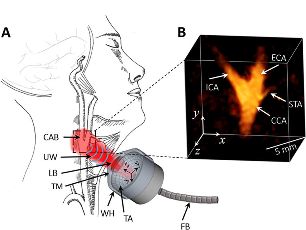
Figure 1. Images show volumetric multispectral optoacoustic tomographic (MSOT) imaging setup. A, Diagram illustrates handheld noninvasive scanning procedure, where volumetric MSOT probe is scanned on skin surface around carotid artery bifurcation area. B, Three-dimensional view of reconstructed volumetric MSOT image of carotid bifurcation captured at video rate of 10 Hz in a 44-year-old man. CAB = carotid artery bifurcation, CCA = common carotid artery, ECA = external carotid artery, FB = fiber bundle, ICA = internal carotid artery, LB = laser beam, STA = superior thyroid artery, TA = transducer array, TM = transparent membrane, UW = ultrasound waves, WH = water holder.
High-res (TIF) version
(Right-click and Save As)
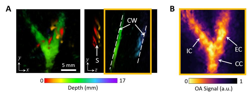
Figure 2. (a, b) Noninvasive volumetric multispectral optoacoustic (OA) tomographic anatomic imaging of carotid artery bifurcation in vivo in a 44-year-old man. (a) Image shows depth characterization. Maximum intensity projections (MIPs) of volumetric reconstructions along z and y directions are color coded to represent depth (in millimeters), where structures in red identify superficial contrast and blue and purple are indicative of deeper structures. (b) MIP of volumetric reconstruction of carotid bifurcation after removal of contrast arising from shallow structures. Orange box in a indicates depth range used for rendering MIP in b. CC = common carotid, CW = carotid wall, EC = external carotid, IC = internal carotid, S = skin.
High-res (TIF) version
(Right-click and Save As)
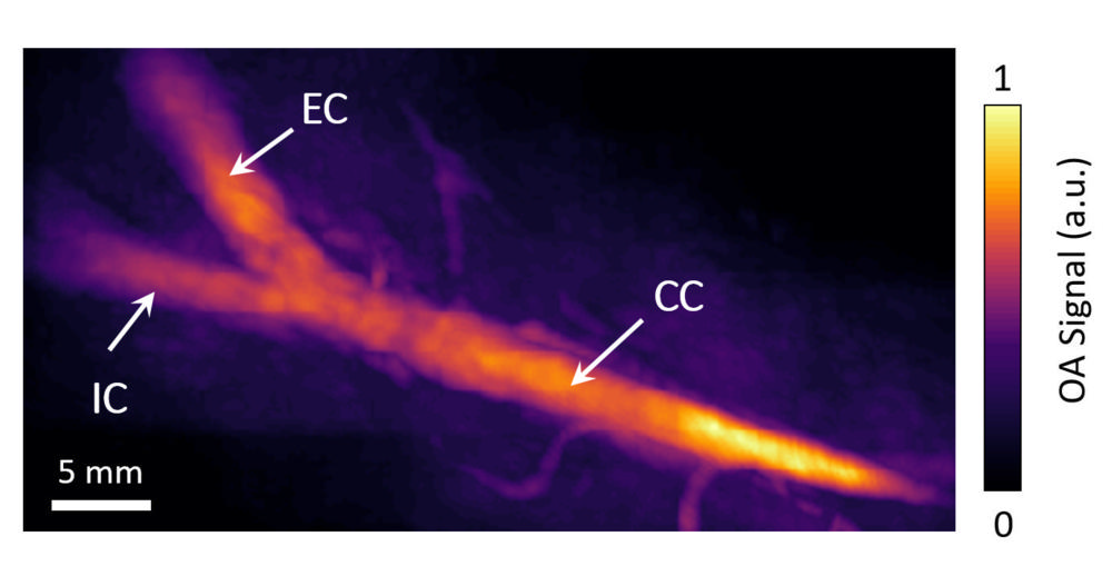
Figure 3. Compounded image of handheld volumetric multispectral optoacoustic (OA) tomography scan along entire carotid artery in a 26-year-old woman. CC = common carotid, EC = external carotid, IC = internal carotid.
High-res (TIF) version
(Right-click and Save As)
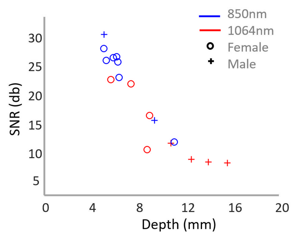
Figure 4. Graph shows signal-to-noise-ratio (SNR) of optoacoustic signal recorded noninvasively from superficial carotid wall and plotted against its depth in 16 volunteers. Measurements were performed at wavelength of either 850 nm (seven women [77.7 percent] with mean age ± standard deviation of 28.28 years ± 2.05; two men [22.22 percent] with mean age of 40.5 years ± 3.5) or 1064 nm (four women [50 percent] with mean age of 27.25 years ± 1.92; four men [50 percent] with mean age of 30 years ± 4.18).
High-res (TIF) version
(Right-click and Save As)
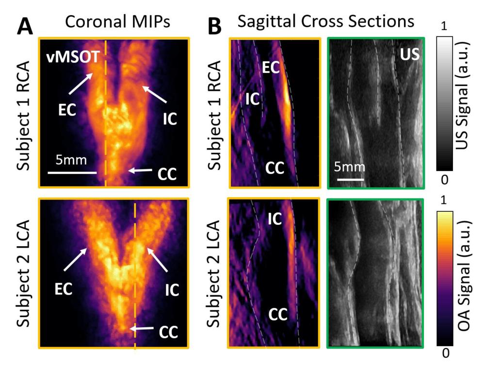
Figure 5. Images show qualitative comparison of image quality between volumetric multispectral optoacoustic tomographic (hereafter, vMSOT) and B-mode US in two volunteers. Right carotid artery (RCA) and left carotid artery (LCA) are shown in a 31-year-old woman (subject 1) and in a 26-year-old woman (subject 2), respectively. A, vMSOT images of carotid artery bifurcation in coronal view represented in maximum intensity projections (MIPs). B, vMSOT and B-mode US images of carotid bifurcation in sagittal cross-sectional views. Orange and green frames correspond to vMSOT and US images, respectively, where orange dashed lines in A indicate section shown in B. CC = common carotid, EC = external carotid, IC = internal carotid, OA = optoacoustic.
High-res (TIF) version
(Right-click and Save As)
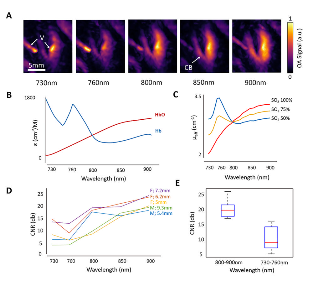
Figure 6. (a) Volumetric multispectral optoacoustic (OA) tomography images show right carotid artery bifurcation in a 23-year-old woman taken in wavelength range of 730–900 nm. (b) Graph shows spectral dependence of optical absorption by oxygenated hemoglobin (HbO) and deoxygenated hemoglobin (Hb). (c) Graph shows simulated wavelength-dependent effective light attenuation coeffi-cient of average soft living tissue. (d) Graph shows contrast-to-noise ratio (CNR) of images as function of wavelength including five volunteers (three of five [60 percent] women with mean age ± standard deviation of 26.6 years ± 3.3; two of five [40 percent] men with mean age of 40.5 years ± 3.5). (e) Graph shows statistical difference of CNR was calculated for longer wavelengths (800–900 nm) and shorter wavelengths (730–760 nm) (P ≤ .001, T test). CB = carotid bifurcation, F = female, M = male, V = vein.
High-res (TIF) version
(Right-click and Save As)