CT Technique Expands Possibilities of Imaging Ancient Remains
Released: September 25, 2018
At A Glance
- The soft tissue in a mummy's hand was successfully imaged down to a microscopic level using phase-contrast CT.
- Researchers were able to see the remains of adipose cells, blood vessels and nerves, and detect blood vessels in the nail bed.
- The findings point the way to a role for phase-contrast CT as a complement to invasive methods used in soft-tissue paleopathology.
- RSNA Media Relations
1-630-590-7762
media@rsna.org - Linda Brooks
1-630-590-7738
lbrooks@rsna.org - Dionna Arnold
1-630-590-7791
darnold@rsna.org
OAK BROOK, Ill. — Researchers in Sweden using computed tomography (CT) have successfully imaged the soft tissue of an ancient Egyptian mummy's hand down to a microscopic level, according to a study published in the journal Radiology.

Non-destructive imaging of human and animal mummies with X-rays and CT has been a boon to the fields of archaeology and paleopathology, or the study of ancient diseases. Imaging studies have contributed to a better knowledge of life and death in ancient times and have the potential to improve our understanding of modern diseases.
Both X-ray and conventional CT take advantage of the fact that materials absorb different amounts of X-rays. This phenomenon, known as absorption contrast, creates different degrees of contrast within an image.
"For studying bone and other hard, dense materials, absorption contrast works well, but for soft tissues the absorption contrast is too low to provide detailed information," said Jenny Romell, M.Sc., from KTH Royal Institute of Technology/Albanova University Center in Stockholm, Sweden. "This is why we instead propose propagation-based phase-contrast imaging."
Propagation-based imaging enhances the contrast of X-ray images by detecting both the absorption and phase shift that occurs as X-rays pass through a sample. The phase effect with X-rays is similar to how a ray of light changes direction as it passes through a lens. Capturing both absorption and phase shift provides higher contrast for soft tissues.
"There is a risk of missing traces of diseases only preserved within the soft tissue if only absorption-contrast imaging is used," Romell said. "With phase-contrast imaging, however, the soft tissue structures can be imaged down to cellular resolution, which opens up the opportunity for detailed analysis of the soft tissues."
Romell and colleagues evaluated phase-contrast CT by imaging a mummified human right hand from ancient Egypt. The hand, today in the collection of the Museum of Mediterranean and Near Eastern Antiquities, was brought to Sweden at the end of the 19th century, along with other mummified body parts and a fragment of mummy cartonnage (papier-mâché case). The cartonnage belonged to an Egyptian man and has been dated to around 400 BCE (before common era). They scanned the entire hand and then performed a detailed scan of the tip of the middle finger.
The estimated resolution of the final images was between 6 to 9 micrometers, or slightly more than the width of a human red blood cell. Researchers were able to see the remains of adipose cells, blood vessels and nerves; they were even able to detect blood vessels in the nail bed and distinguish the different layers of the skin.
"With phase-contrast CT, ancient soft tissues can be imaged in a way that we have never seen before," Romell said.
The findings point the way to a role for phase-contrast CT as an adjunct or alternative to invasive methods used in soft-tissue paleopathology that require extraction and chemical processing of the tissue. Due to their potentially destructive nature, these methods are undesirable or unacceptable for the analysis of many old and fragile specimens.
"Just as conventional CT has become a standard procedure in the investigation of mummies and other ancient remains, we see phase-contrast CT as a natural complement to the existing methods," Romell said. "We hope that phase-contrast CT will find its way to the medical researchers and archaeologists who have long struggled to retrieve information from soft tissues, and that a widespread use of the phase-contrast method will lead to new discoveries in the field of paleopathology."
"Soft-Tissue Imaging in a Human Mummy: Propagation-based Phase-contrast CT." Collaborating with Romell were William Vågberg, M.Sc., Mikael Romell, M.D., Sofia Häggman, Ph.D., Salima Ikram, Ph.D., and Hans M. Hertz, Ph.D.
Radiology is edited by David A. Bluemke, M.D., Ph.D., University of Wisconsin School of Medicine and Public Health, Madison, Wis., and owned and published by the Radiological Society of North America, Inc. (http://radiology.rsna.org/)
RSNA is an association of over 54,200 radiologists, radiation oncologists, medical physicists and related scientists, promoting excellence in patient care and health care delivery through education, research and technologic innovation. The Society is based in Oak Brook, Ill. (RSNA.org)
For patient-friendly information on CT, visit RadiologyInfo.org.
Videos and Images
Video 1. Axial reconstruction of mummified hand. Animation through the full stack of axial sections from the overview tomography of the mummy hand.
Download MP4
(Right-click and Save As)
Video 2. Axial reconstruction of fingertip. Animation through stack of axial sections from the high-resolution tomography of the fingertip.
Download MP4
(Right-click and Save As)
Video 3. Virtual sectioning of fingertip (coronal). Animation through virtual coronal sections of the stack in Video 2.
Download MP4
(Right-click and Save As)
Video 4. Virtual sectioning of fingertip (sagittal). Animation through virtual sagittal sections of the stack in Video 2.
Download MP4
(Right-click and Save As)
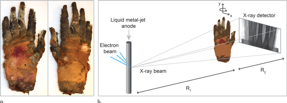
Figure 1. Images show propagation-based phase-contrast CT of mummified hand. (a) Photograph of human hand that was imaged shows palmar (left) and dorsal (right) views. Hand and parts of fingers are wrapped in linen. Most of skin and all fingernails are well preserved. (b) Schematic of experimental arrangement shows microfocus x-ray source, sample placed on rotation stage, and x-ray detector.
High-res (TIF) version
(Right-click and Save As)
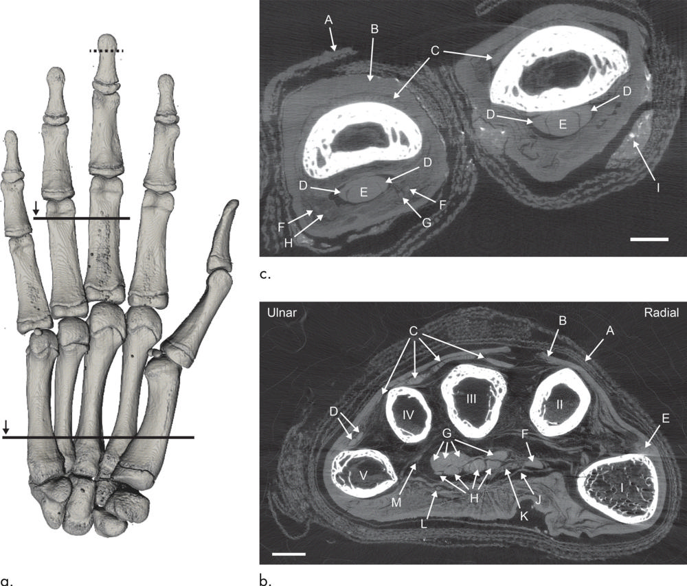
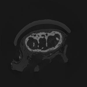
Figure 2. Images show CT of mummified hand. (a) Palmar view of volume rendering depicts bones. (b) Axial view of hand. Arrows indicate linen (A), skin (B), tendons of extensor digitorum communis (C), tendons of extensor digiti minimi (D), tendons of extensor pollicis longus (E), tendons of flexor pollicis longus (F), tendons of flexor digitorum profundus (G), tendons of flexor digitorum superficialis (H), distal portion of carpal ligament (J), median nerve (K), ulnar nerve and/or superficial branch of ulnar artery (L), and possibly deep branch of ulnar artery (M). Scale bar represents 5 mm. (c) Axial section through proximal phalanges of third and fourth fingers. Arrows indicate linen (A), skin (B), tendons of extensor digitorum (C), tendons of flexor digitorum superficialis (D), tendons of flexor digitorum profundus (E), proper palmar digital arteries (F), proper palmar digital nerves of median nerve (G) and of ulnar nerve (H), and residues from embalming process (I).
High-res (TIF) version
(Right-click and Save As)
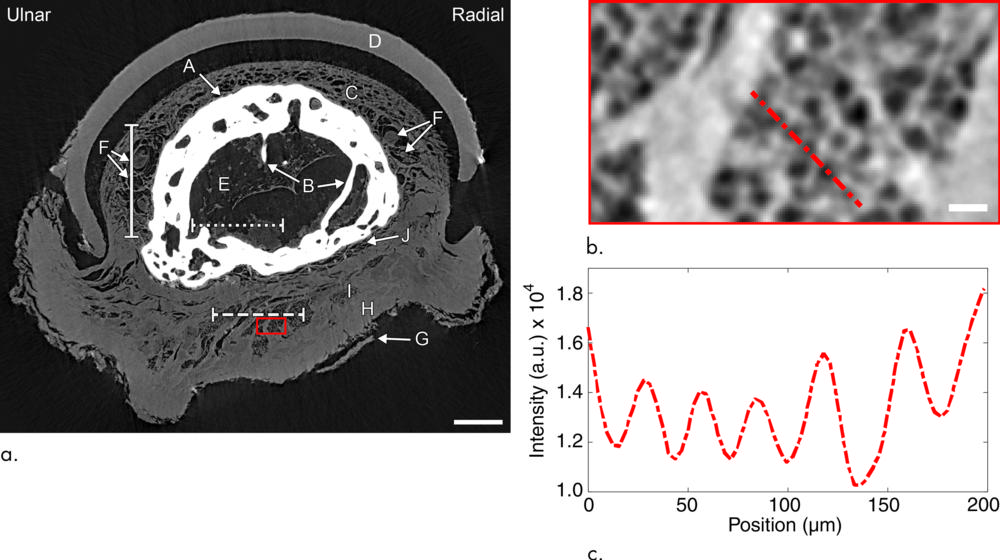
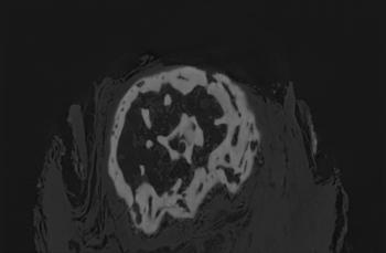
Figure 3. Images show fingertip of mummified hand. (a) Axial section through distal phalange of middle finger. Arrows indicate bone (A) with trabeculae (B), nail bed (C), nail (D), bone marrow (E), vessels and/or nerves (F), epidermis (G), dermis (H), hypodermis (I), and periosteum (J). Sections at the three lines are shown in Figure 4. (b) Detailed view of hypodermis. (c) Line profile shows intensity along dashed red line in b.
High-res (TIF) version
(Right-click and Save As)
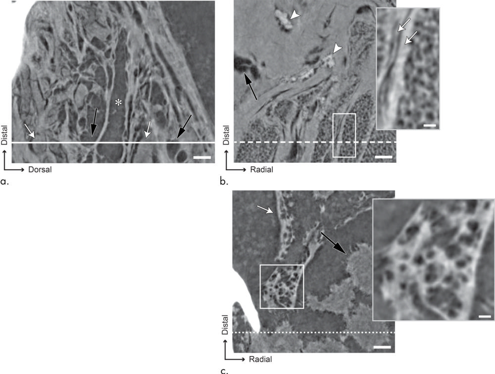
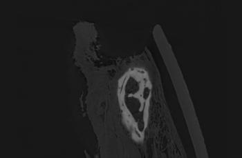
Figure 4. (a-c) Images show soft-tissue microanatomy of fingertip with sections at solid, dashed, and dotted lines, respectively, from Figure 3a. (a) Image shows vessels and possibly nerves. (*) indicates hollow vessel. Black arrows indicate neighboring hollow structurers, and white arrows show structures of similar shapes, but filled. (b) Image shows hypodermis. Black arrow indicates air pocket. Arrowheads indicate dense, so far unidentified structures. Inset shows enlarged view of adipose tissue, where remains of cellular structures can be seen. Arrows mark connective tissue septa. (c) Images depict two types of materials: dehydrated bone marrow (white arrow) and one agglomeration, possibly residue from mummification process (black arrow). Cavities in bone marrow are shown in inset.
High-res (TIF) version
(Right-click and Save As)
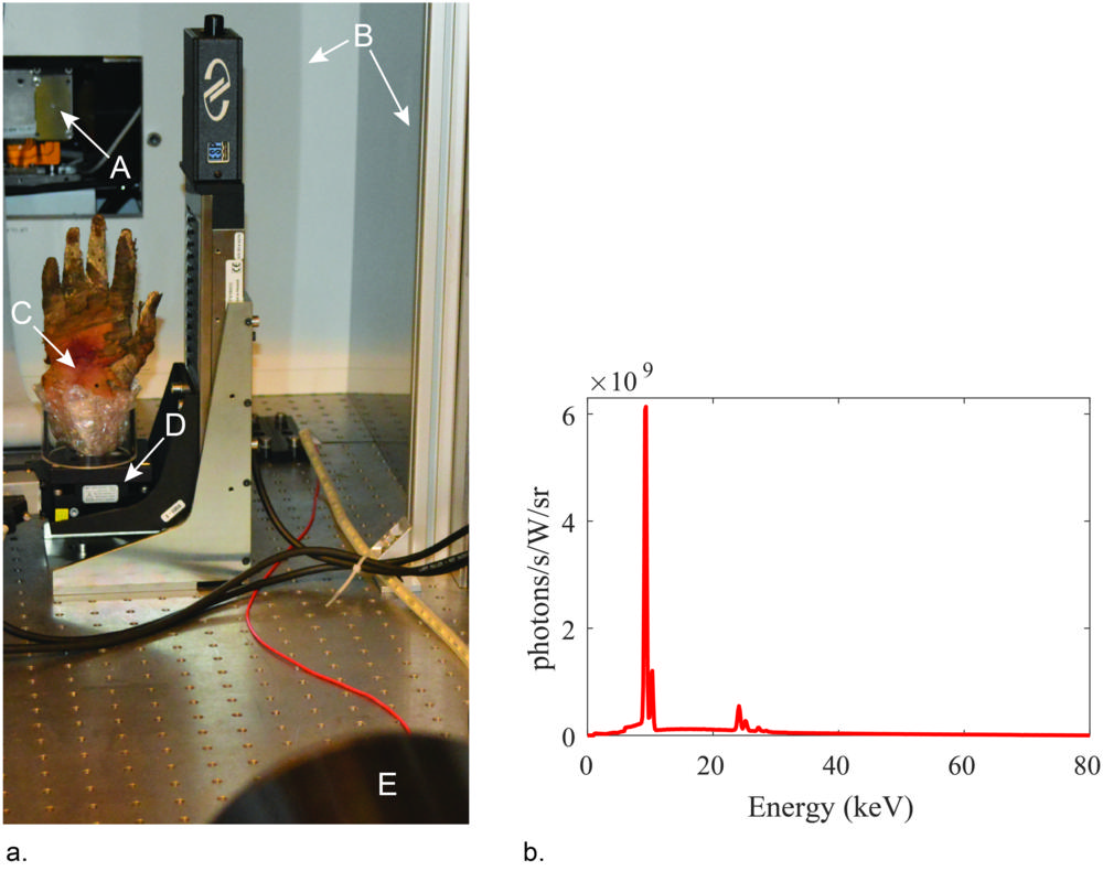
Figure 5. (a) Photograph of the phase-contrast imaging arrangement as viewed from the detector: (A) x-ray source with output window, (B) radiation shielding around source and experimental arrangement, (C) sample placed in a plastic cylinder and padded with plastic foam, (D) rotation stage and (E) an x-ray camera. (b) Spectrum of the liquid-metal-jet x-ray source measured at acceleration voltage 80 kV.
High-res (TIF) version
(Right-click and Save As)
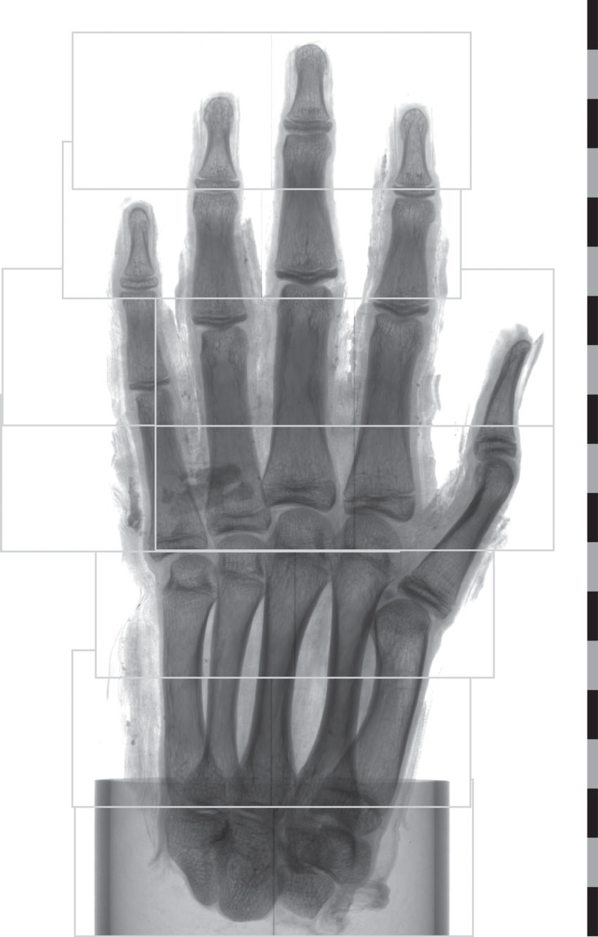
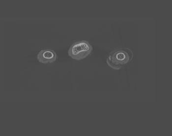
Figure 6. Regions for tomographic scans. For the imaging of the hand, a total of nine tomographic scans were performed. Here, the field of view for each scan is shown. Before tomographic reconstruction, the left regions of the fourth and fifth scan from the bottom were stitched together with their respective right counterpart. The scale shows centimeters.
High-res (TIF) version
(Right-click and Save As)