MRI May Predict Neurological Outcomes for Cardiac Arrest Survivors
Released: October 18, 2017
At A Glance
- Using advanced MRI techniques, researchers may be able to predict neurological outcomes for survivors of cardiac arrest.
- Researchers assessed the brain functional connectivity in 46 patients who were in a coma following cardiac arrest.
- The results showed that connectivity measures could be predictors of long-term recovery potential in patients with cardiac arrest-related brain damage.
- RSNA Media Relations
1-630-590-7762
media@rsna.org - Linda Brooks
1-630-590-7738
lbrooks@rsna.org - Dionna Arnold
1-630-590-7791
darnold@rsna.org
OAK BROOK, Ill. – MRI-based measurements of the functional connections in the brain can help predict long-term recovery in patients who suffer neurological disability after cardiac arrest, according to a study appearing online in the journal Radiology.

Cardiac arrest, or abrupt loss in heart function, is a common and often deadly occurrence that affects hundreds of thousands of people every year in the United States alone, according to the American Heart Association. Many patients who survive end up with severe neurological disabilities, as the temporary loss of oxygenated blood flow to the brain can result in widespread neuronal cell death.
“Current methods to predict future levels of function for these survivors have limited accuracy,” said study lead author Robert D. Stevens, M.D., from Johns Hopkins University School of Medicine in Baltimore. “We need better methods to help clinicians understand the magnitude of these injuries and make more accurate predictions on recovery, thereby enabling more informed decision-making.”
For the study, Dr. Stevens and colleagues used advanced MRI techniques like diffusion tensor imaging and resting-state functional MRI (fMRI) to focus on the brain’s large-scale functional integration. This “network of networks,” or connectome, represents the ensemble of different neuronal populations in the brain that work together to perform tasks.
The researchers assessed the brain’s functional connectivity in 46 patients who were in a coma following cardiac arrest. The imaging, performed within two weeks of cardiac arrest, included studies of brain structure and function. Functional imaging focused on four well-characterized networks in the brain, including the default mode network, which is active when a person is not engaged in a specific task, and the salience network, a collection of brain regions that select which stimuli are deserving of our attention.
One year after the patients’ cardiac arrests, the researchers assessed the patients with the Cerebral Performance Category Scale, a commonly used measure of neurological function following cardiac arrest. Eleven patients had favorable outcomes. Functional connectivity was stronger in those who achieved higher levels of independence at one year compared with those who were heavily dependent. The changes in functional connectivity between networks predicted outcomes with greater accuracy than any of the MRI structural measures tested.
“This is game-changing information about what happens in the brains of people who suffer cardiac arrest,” Dr. Stevens said. “We realize that network architectures can be selectively disrupted in this setting.”
A key predictor of outcomes was the interaction between the brain’s default mode and salience networks. These two networks are normally anti-correlated, meaning that as the default mode network becomes more active, activity is reduced in the salience network, and vice versa. When researchers compared the brain imaging results of patients who had favorable outcomes with those who did not, they noticed a stark difference.
“Anti-correlation was preserved in patients who recovered and abolished in those who did not,” Dr. Stevens said. “Relative preservation of this anti-correlation was the most robust signal of a favorable outcome.”
The results indicate that connectivity measures could be early markers of long-term recovery potential in patients with cardiac arrest-related brain damage, the researchers said.
While researchers don’t expect connectome analysis with MRI to be the single “magic bullet” solution to predicting outcomes, it could increase the confidence that clinicians have in communicating with patients’ families in the wake of cardiac arrest. Additionally, fMRI could aid in the development of therapeutic interventions for neurologically disabled patients.
“Connectome studies have the potential to change not only outcome prediction, but to guide treatment as well,” Dr. Stevens said.
“Early Functional Connectome Integrity and 1-Year Recovery in Comatose Survivors of Cardiac Arrest.” Collaborating with Dr. Stevens were Haris I. Sair, M.D., Yousef Hannawi, M.D., Shanshan Li, Ph.D., Joshua Kornbluth, M.D., Athena Demertzi, Ph.D., Carol Di Perri, M.D., Ph.D., Russell Chabanne, M.D., Betty Jean, M.D., Habib Benali, Ph.D., Vincent Perlbarg, Ph.D., James Pekar, Ph.D., Charles-Edouard Luyt, M.D., Ph.D., Damien Galanaud, M.D., Ph.D., Lionel Velly M.D., Louis Puybasset M.D., Ph.D., Steven Laureys, M.D., Ph.D., and Brian Caffo, Ph.D., on behalf of the NeuroImaging for Coma Emergence and Recovery (NICER) Consortium.
Radiology is edited by Herbert Y. Kressel, M.D., Harvard Medical School, Boston, Mass., and owned and published by the Radiological Society of North America, Inc. (http://radiology.rsna.org/)
RSNA is an association of over 54,600 radiologists, radiation oncologists, medical physicists and related scientists, promoting excellence in patient care and health care delivery through education, research and technologic innovation. The Society is based in Oak Brook, Ill. (RSNA.org)
For patient-friendly information on brain MRI, visit RadiologyInfo.org.
Images (.JPG and .TIF format)
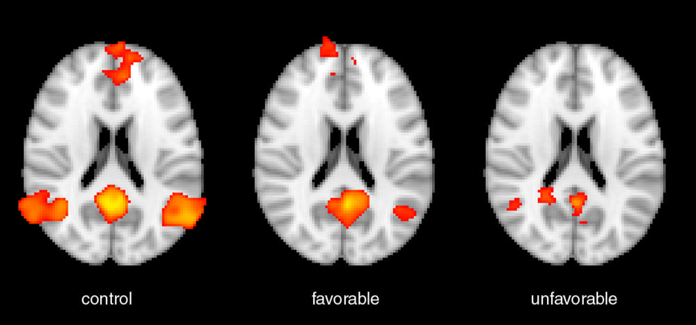
Figure 1. Functional MRI activation (shown as red-orange) as seen in a representative healthy control subject (left), a cardiac arrest patient who had a good functional outcome (middle), and a cardiac arrest patient who had a poor functional outcome (right). The formation shown here, called the default mode network, loses internal coherence (connectivity) in cardiac arrest patients, and the magnitude of that loss is inversely proportional to probability of recovery.
High-res (TIF) version
(Right-click and Save As)
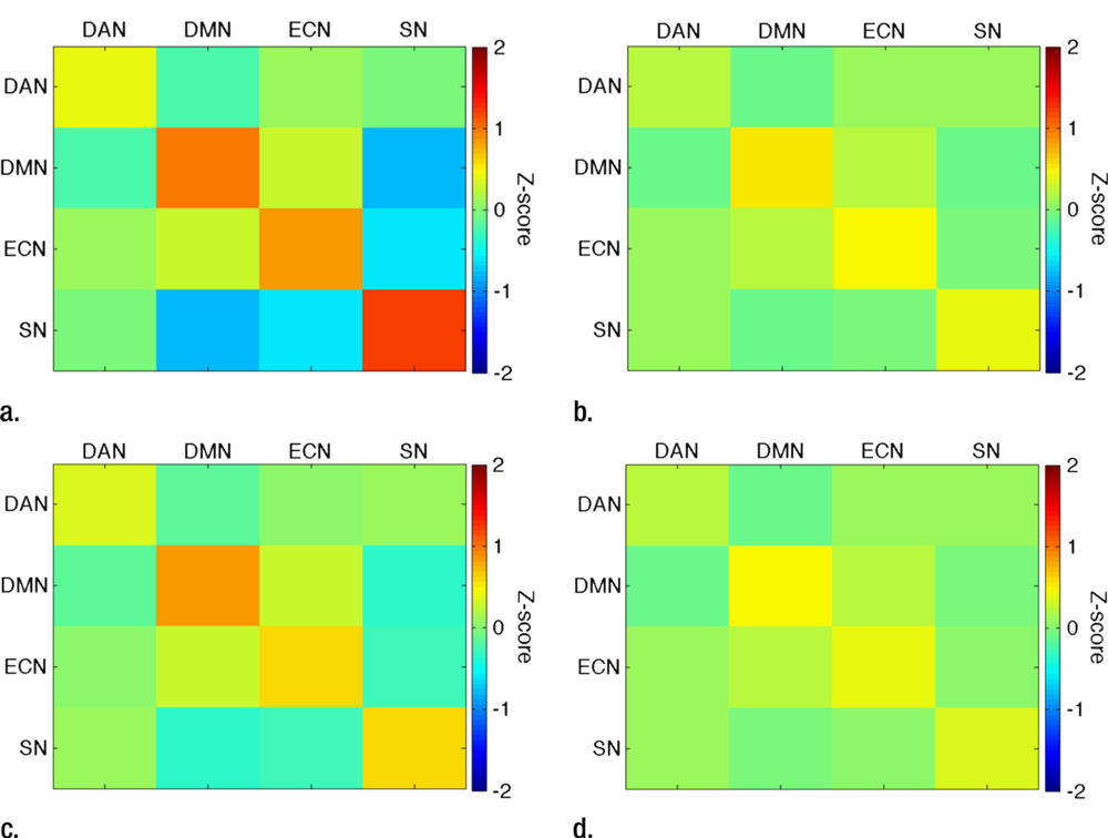
Figure 2. This figure displays the strength of functional MRI connectivity within and between networks of the brain. Warmer colors (yellow-orange-red) indicate positive correlations, colder colors (shades of blue) indicate negative correlations, and green indicates absence of correlation. The connectivity seen in healthy controls (a) is reduced in cardiac arrest patients (b), and this reduction is more pronounced in cardiac arrest patients who had a poor functional outcome (d) when compared to those who had a poor functional outcome (c). Acronyms denote individual networks (DAN=dorsal attention network. DMN=default mode network. ECN=executive control network. SN=salience network).
High-res (TIF) version
(Right-click and Save As)
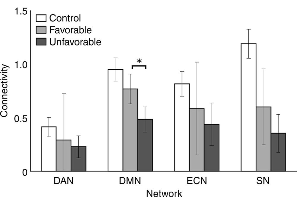
Figure 3. This plot compares the connectivity within networks in healthy controls (white) which is reduced in cardiac arrest patients who had a good functional outcome (light grey), and even further reduced in those with a poor functional outcome (dark grey). Acronyms denote individual networks (DAN=dorsal attention network. DMN=default mode network. ECN=executive control network. SN=salience network).
High-res (TIF) version
(Right-click and Save As)
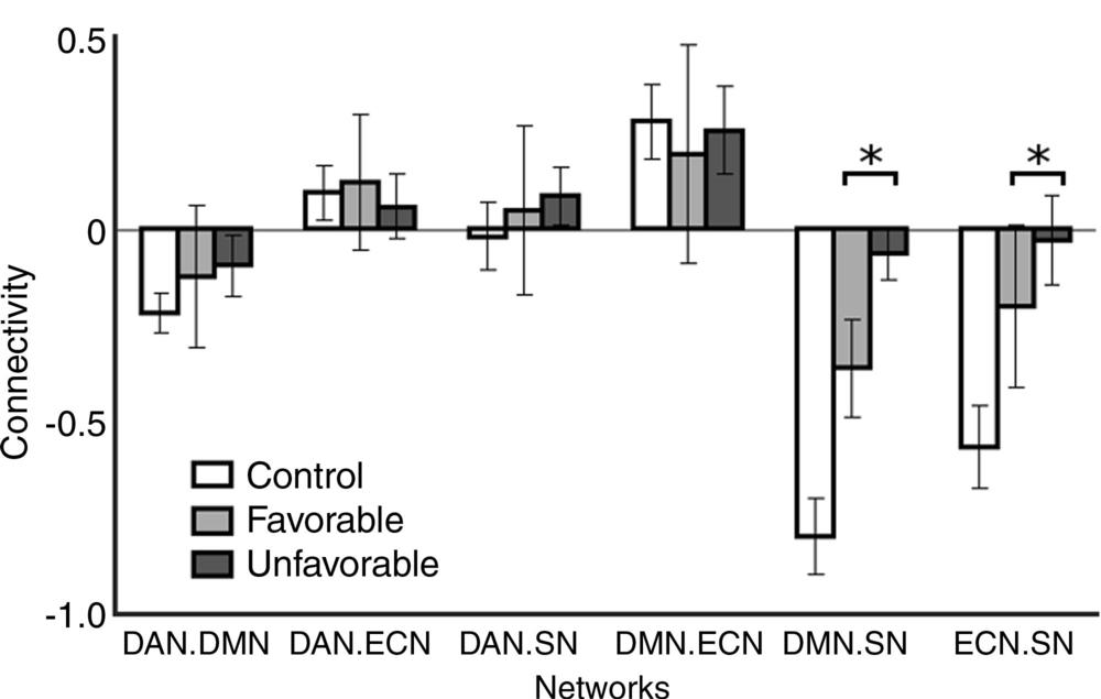
Figure 4. This plot compares the connectivity between networks in healthy controls (white) which is reduced in cardiac arrest patients who had a good functional outcome (light grey), and even more reduced in those with a poor functional outcome (dark grey). Negative values indicate inverse correlation (anticorrelation) between networks. A notable finding in our study was that the inverse correlations between the salience network (SN) and two other networks were the best predictors of one year functional outcome. Acronyms denote network pairs (DAN.DMN=connectivity between dorsal attention network and default mode network. DAN.ECN=connectivity between dorsal attention network and executive control network. DAN.SN=connectivity between dorsal attention network and salience network. DMN.ECN=connectivity between default mode network and executive control network. DMN.SN= connectivity between default mode network and salience network. ECN.SN= connectivity between executive control network and salience network).
High-res (TIF) version
(Right-click and Save As)
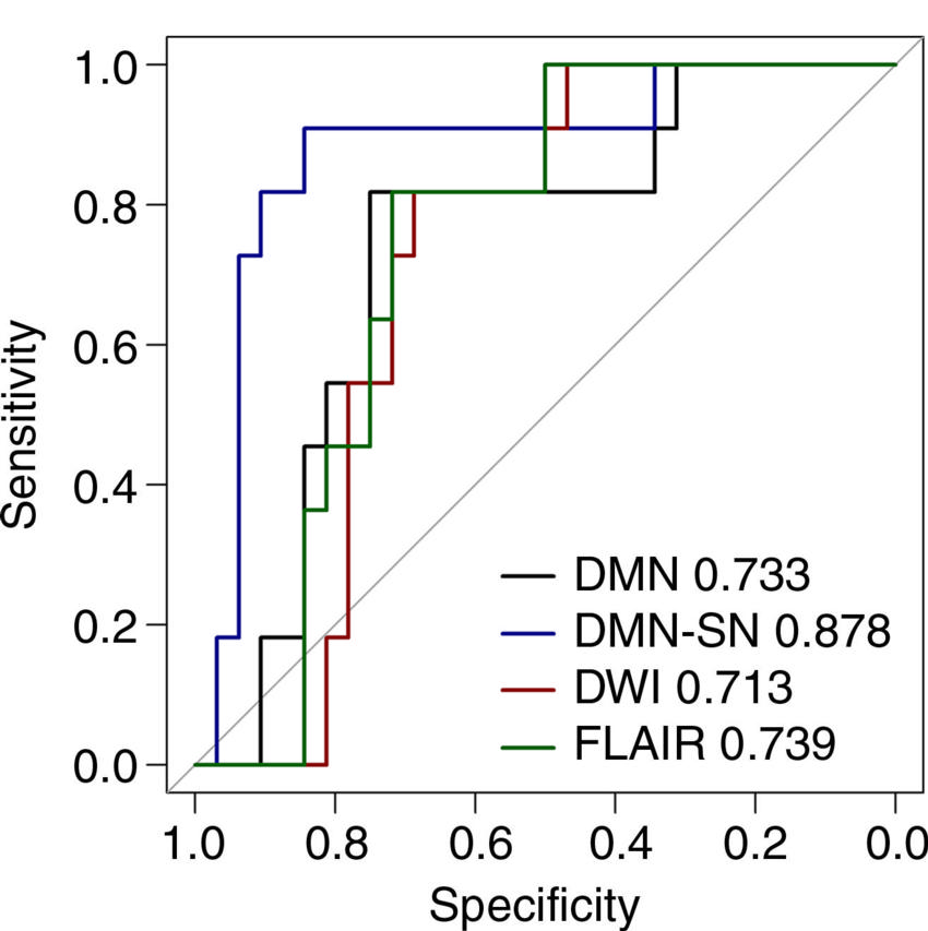
Figure 5. The family of curves shown here contrasts the ability of different MRI-based functional and structural variables to predict one year functional outcome. The area under the curves is a measure of the discriminative ability of each variable. Here, it is shown that the inverse correlation between the salience network and the default mode network (DMN-SN) is the strongest predictor, better than the internal connectivity of the default mode network, better also than the signal changes detected using structural MRI (FLAIR, DWI).
High-res (TIF) version
(Right-click and Save As)