Imaging Predicts Long-Term Effects in Veterans with Brain Injury
Released: March 29, 2016
At A Glance
- Researchers used DTI to assess the long-term effects of combat-induced brain injury in military veterans.
- DTI measurements correlated with clinical outcomes, including symptom severity and healthcare utilization.
- Loss of white matter integrity in the brain has a direct, measurable effect on clinical outcomes in veterans with MTBI.
- RSNA Media Relations
1-630-590-7762
media@rsna.org - Linda Brooks
1-630-590-7738
lbrooks@rsna.org
OAK BROOK, Ill. — Diffusion tensor imaging (DTI), a type of MRI, may be able to predict functional post-deployment outcomes for veterans who sustained mild traumatic brain injury (MTBI), or concussion, during combat, according to a new study published in the journal Radiology.

Current assessment of MTBI remains challenging due to the difficulties in establishing the diagnosis, predicting outcomes and separating the effects of MTBI from other conditions like post-traumatic stress disorder (PTSD).
DTI uses measurements of water movement in the brain to detect abnormalities, particularly in white matter. Previous studies have linked DTI metrics to neurocognitive function and short-term functional outcomes in groups of patients. The desire to uncover possible long-term effects spurred Jeffrey B. Ware, M.D., from the Philadelphia VA Medical Center in Philadelphia, Pa., to evaluate combat veterans using this technique.
Dr. Ware and colleagues used brain MRI and DTI to study 57 military veterans who had a clinical diagnosis of MTBI upon return from deployment. The average length of time between injury and post-deployment evaluation was 3.8 years with an average follow-up duration of 1.4 years.
"All conventional MR images were interpreted as normal," Dr. Ware said. "We retrospectively analyzed the data from the DTI sequence to derive measures of white matter integrity, which we compared to clinical measures and subsequent outcome measures 6 months to 2.5 years after the initial evaluation."
The results showed significant associations between initial post-deployment DTI measurements and neurobehavioral symptoms, timing of injury, and subsequent functional outcomes. The measurements also correlated with greater healthcare utilization among veterans with MTBI.
Following initial post-deployment evaluation, 34 of the study participants returned to work. Veterans who did not return to work displayed significantly lower fractional anisotropy (FA) and higher diffusivity in a specific brain region, the left internal capsule. These measures imply less structural integrity in that area of the brain. As this region is known to contain important fibers providing motor stimulation to the typically dominant right side of the body, the results may provide a correlation between impairments in fine motor functioning and inability to return to work.
"Our findings suggest that differences in white matter microstructure may partially account for the variance in functional outcomes among this population. In particular, loss of white matter integrity has a direct, measurable effect," Dr. Ware said. "It was illuminating to see the association between measures of white matter integrity and important outcomes occurring months to years down the road in our study population."
"Combat-related Mild Traumatic Brain Injury: Association between Baseline Diffusion-Tensor Imaging Findings and Long-term Outcomes." Collaborating with Dr. Ware on this paper were Rosette C. Biester, Ph.D., Elizabeth Whipple, M.S., Keith M. Robinson, M.D., Richard J. Ross, M.D., Ph.D., and Paolo G. Nucifora, M.D., Ph.D.
Radiology is edited by Herbert Y. Kressel, M.D., Harvard Medical School, Boston, Mass., and owned and published by the Radiological Society of North America, Inc. (http://radiology.rsna.org/)
RSNA is an association of more than 54,000 radiologists, radiation oncologists, medical physicists and related scientists, promoting excellence in patient care and health care delivery through education, research and technologic innovation. The Society is based in Oak Brook, Ill. (RSNA.org)
For patient-friendly information on MRI, visit RadiologyInfo.org.
Images (.JPG and .TIF format)
and Video clips (.mp4 format)
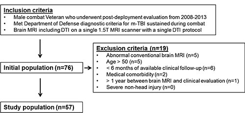
Figure 1. Overview of study population, inclusion criteria, and exclusion criteria. m-TBI = mild TBI.
High-res (TIF) version
(Right-click and Save As)
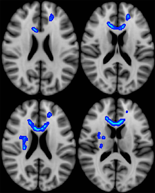
Figure 2. Blue indicates regions of the brain in which lower fractional anisotropy (a measure of microstructural integrity) correlated with more severe neurobehavioral symptoms. Veterans with the most severe symptoms had lower microstructural integrity in these regions.
High-res (TIF) version
(Right-click and Save As)
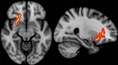
Figure 3. Red/yellow indicates regions of the brain in which fractional anisotropy correlated with time since most severe TBI event. Veterans with more recent injury had lower microstructural integrity in these regions.
High-res (TIF) version
(Right-click and Save As)
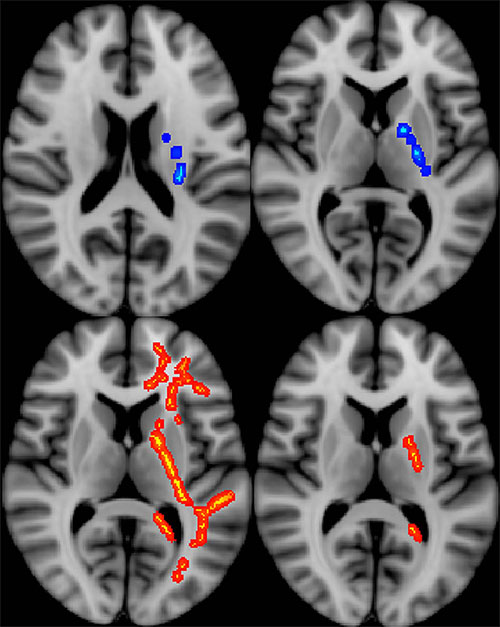
Figure 4. All colored regions signify regions of the brain in which different diffusion- tensor imaging measures depict lower microstructural integrity in Veterans who did not return to work after returning from deployment, during the study period.
High-res (TIF) version
(Right-click and Save As)
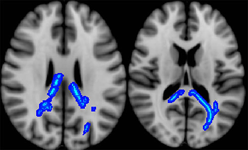
Figure 5. Blue indicates regions of the brain in which lower fractional anisotropy correlated with greater healthcare utilization (more frequent healthcare visits). Veterans who had higher utilization over the study period had lower microstructural integrity in these regions.
High-res (TIF) version
(Right-click and Save As)